Search results (16 results)
-
 Angle Closure Glaucoma
Nov 14 2019 by Jennifer Schiefer, CRA, COA
96-year-old female who presented in clinic with eye pain x 3 weeks and HM Va. Unique case in pseudophakic eye. Exam showed sectoral area of NVI. Questionable NVG after RVO.
Photographer: Jennifer Schiefer
Condition/keywords: angle closure, anterior chamber, neovascular glaucoma
-
 Angle Closure/ UBM and Gonioscopy
Jul 8 2013 by Jason S. Calhoun
Patient came in a day after dilation with severe headache and nausea. Patient's IOP was 64 in the right eye. Patient ended up having angle closure attack. Promoted severe nausea and eye pain. Peripheral iridotomy was performed and IOP dropped slowly. Patient was followed up the next morning and will be seen 2-days after that.
Photographer: Jason S. Calhoun, Department of Ophthalmology, Mayo Clinic Jacksonville, Florida
Condition/keywords: angle closure, gonioscopy, ultrasound
-
 Iridocorneal Angle
Sep 13 2013 by Maria Ana Martinez-Castellanos, MD
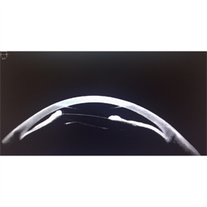
Angle OCT of a right eye of a 4 months old male patient with retinopathy of prematurity stage 5.
Photographer: Maria A. Martinez-Castellanos. Asociación para Evitar la Ceguera en Mexico
Condition/keywords: angle closure, retinopathy of prematurity (ROP), retinopathy of prematurity, stage 5
-
 pupillary block; periph uveitis
Feb 14 2013 by From the Collections of Thomas M. Aaberg, MD and Thomas M. Aaberg Jr., MD
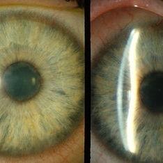
diffuse and slit-beam anterior segment photographs showing pupillary-block angle closure associated with uveitis. pupillary block; angle closure; uveitis; posterior synechiae
Condition/keywords: angle closure, posterior synechiae, pupillary block, uveitis
-
 UBM Angle Closure
Jul 14 2013 by Jason S. Calhoun
UBM ultrasound shows angle closure. Proceeded with PI to open angle.
Photographer: Jason S. Calhoun, Department of Ophthalmology, Mayo Clinic Jacksonville, Florida
Imaging device: UBM
Condition/keywords: angle closure
-
 Angle Closure Glaucoma
Jul 29 2013 by H. Michael Lambert, MD
Angle closure glaucoma.
Condition/keywords: acute angle-closure glaucoma
-
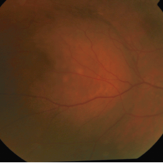
 Choroidal Metastasis
May 31 2016 by MAHTA DOUSTKHAH
Fundus photograph of a 62-year-old Indian lady with choroidal metastasis secondary to primary lung adenocarcinoma (presented with left angle closure glaucoma- but left eye no fundus view).
Photographer: Mahta Doustkhahvajari, University of Malaya
Condition/keywords: choroidal metastasis
-
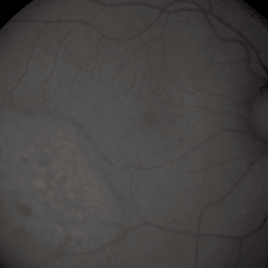 Choroidal Metastasis
May 31 2016 by MAHTA DOUSTKHAH
Fundus photograph (red -free) of a 62-year- old Indian lady with choroidal metastasis secondary to primary lung adenocarcinoma (presented with left angle closure glaucoma- but left eye no fundus view).
Photographer: Mahta Doustkhahvajari, University of Malaya
Condition/keywords: choroidal metastasis
-
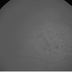 Choroidal Metastasis
May 31 2016 by MAHTA DOUSTKHAH
Fundus photograph (red -free) of a 62-year- old Indian lady with choroidal metastasis secondary to primary lung adenocarcinoma (presented with left angle closure glaucoma- but left eye no fundus view).
Photographer: Mahta Doustkhahvajari, University of Malaya
Condition/keywords: choroidal metastasis
-
 Glaukomflecken
Oct 23 2017 by Claire Kiernan, MD
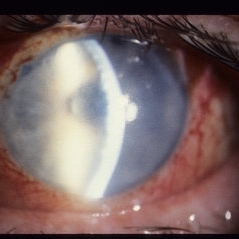
Slit lamp photograph of a 59-year-old man with recent-onset severe eye pain noted to have glaukomflecken consistent with recent episode of angle closure glaucoma.
Photographer: Steve Moser, University of Tennessee Hamilton Eye Institute; Joe Mastellone, University of Tennessee Hamilton Eye Institute
Condition/keywords: angle-closure glaucoma interval, glaucoma anterior segment anomalies
-
 Multiple Ciliary Body Cysts
Aug 7 2020 by Rinal Pandit
Ultrasound biomicroscopy image (Transverse scan) of a 30-year-old female with occludable angle. For all young patients presenting with angle closure ,UBM should preferably be performed. It helps establish the cause of narrowing , such as a pseudoplateau configuration in this case produced by multiple CB cysts.
Photographer: Dr Rinal Pandit, Raghudeep eye hospital, Ahmedabad
Imaging device: UBM, ABsolu, Quantel Medical
Condition/keywords: ciliary body cyst
-
 Ophthalmic Artery Occlusion
Dec 17 2018 by Evan Frigoletto
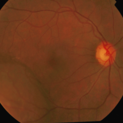
Male patient with known left-sided 100% carotid artery occlusion presents with ipsilateral nonperfusion of the ophthalmic artery. Presenting visual acuity is NLP, with normal IOP. Fundus photograph shows severely attenuated arteries and veins with diffuse peripheral retinal atrophy and disc pallor. Other ocular findings consistent with ischemia include diffuse conjunctival injection, florid iris neovascularization and 360 degree synechial angle closure.
Photographer: Diane Zellers, Geisinger Eye Institute, Danville, Pennsylvania
Imaging device: Montage photograph with Zeiss FF450 plus
Condition/keywords: ophthalmic artery occlusion
-
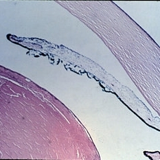 Slide 10-17
Feb 26 2019 by Lancaster Course in Ophthalmology
Angle-closure glaucoma. Histologic section ( x 16). The lens is excessively large and presses against the pupil, resulting in pupillary block, iris bombe, and angle closure. (Scheie Eye Institute, No. 5879.)
Condition/keywords: angle-closure glaucoma interval
-
 Suprachoroidal Hemorrhage
Sep 2 2020 by Rinal Pandit
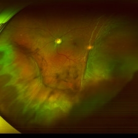
Fundus photograph of left eye of a 56-year-old female with primary angle closure glaucoma showing massive hemorrhagic choroidal detachment that developed following trabeculectomy surgery. Suprachoroidal hemorrhage is defined as the accumulation of blood within the potential space between the choroid and sclera, with the source of the blood being the long or short posterior ciliary artery. Delayed suprachoroidal hemorrhage (DSHC) remains one of the most dreaded and sight threatening complications of glaucoma filtration surgery. The risk factors include old age, hypertension, high myopia, arteriosclerosis, chronically elevated IOP, sudden hypotony, trauma, aphakia/pseudophakia, prior vitrectomy, history of 5 FU injections and anti-platelet agents. The incidence of postoperative SCH after trabeculectomy varies between 0.6%- 1.4%. DSCH after surgery varies considerably in severity but is generally characterized by the sudden onset of severe pain, decreased vision, and a shallow anterior chamber usually associated with raised intraocular pressure. B-scan ultrasonography can help to distinguish serous from hemorrhagic choroidals.Suprachoroidal hemorrhages appear as dome-shaped elevations of the retina with increased echo densities that are often heterogeneous and within the suprachoroidal space. Choroidal effusions appear as dome-shaped elevations with hypoechoic suprachoroidal space. The first step in the management is the timely diagnosis. Medical management includes oral and topical antiglaucoma drugs to lower IOP, oral and topical steroids to control inflammation and topical cycloplegics and oral analgesics to tackle pain. Serial ultrasound B scans of the affected eye should be performed in order to monitor progression of the SCH and help determine apposition, height, and liquefaction of the SCH. Indications of surgical drainage include non resolution with medical management,concurrent retinal detachment, central retinal apposition (kissing choroidals) and incarceration of vitreous in the wound site. The ideal time of drainage is between 7-14 days depending upon clot lysis. The prognosis of both intraoperative and postoperative SCH is poor. An overwhelming majority of patients do not achieve pre-hemorrhage visual acuity and most do not recover to a visual acuity of 20/200 or better. The major determinants of good or bad visual outcomes of SCH’s are preoperative visual acuity and retinal detachment at the time of hemorrhage, respectively.
Imaging device: OPTOS,Ultra wide field retinal imaging system
Condition/keywords: suprachoroidal hemorrhage, trabeculectomy, ultra-wide field imaging
-
 Uveal Effusion Syndrome in a Nanophthalmic Eye
Apr 3 2025 by Gustavo Uriel Fonseca Aguirre
Ultrasound biomicroscopy longitudinal section of a nanophthalmic eye demonstrating a shallow anterior chamber, angle closure, ciliary body edema, and supraciliary and sub-Tenon fluid accumulation.
Photographer: Gustavo U. Fonseca Aguirre, Hospital Conde de Valenciana, Ciudad de México
Condition/keywords: nanophthalmos, uveal effusion syndrome
 Loading…