 Initializing download.
-
 By Evan Frigoletto
Geisinger Commonwealth School of Medicine
Co-author(s): Dr. Gordon Crabtree, Dr. Benjamin Hale, Geisinger Eye Institute, Danville PA - Uploaded on Dec 17, 2018.
- Last modified by Caroline Bozell on Dec 18, 2018.
- Rating
- Appears in
- Miscellaneous
- Condition/keywords
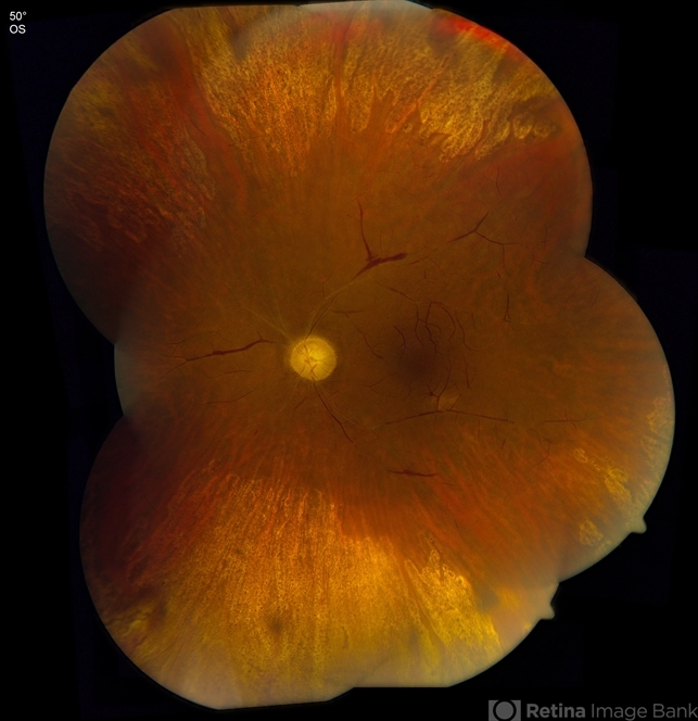
- ophthalmic artery occlusion
- Photographer
- Diane Zellers, Geisinger Eye Institute, Danville, Pennsylvania
- Imaging device
- Montage photograph with Zeiss FF450 plus
- Description
- Male patient with known left-sided 100% carotid artery occlusion presents with ipsilateral nonperfusion of the ophthalmic artery. Presenting visual acuity is NLP, with normal IOP. Fundus photograph shows severely attenuated arteries and veins with diffuse peripheral retinal atrophy and disc pallor. Other ocular findings consistent with ischemia include diffuse conjunctival injection, florid iris neovascularization and 360 degree synechial angle closure.


