Search results (38 results)
-
 Retinitis Pigmentosa (RP)
Sep 21 2023 by Ben Serar
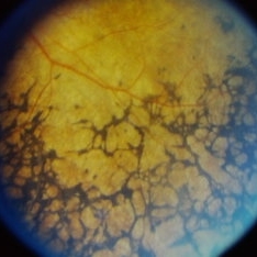
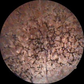
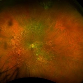
Fundus photograph showing bony spicules and arteriolar attenuation in a case of Retinitis Pigmentosa.
Condition/keywords: Retinitis Pigmentosa (RP)
-
 Retinitis Pigmentosa (RP)
Sep 21 2023 by Ben Serar
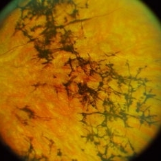
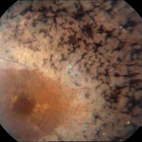
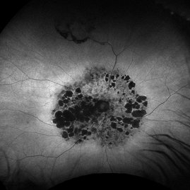
Fundus photograph showing bony spicules in a case of Retinitis Pigmentosa.
Condition/keywords: Retinitis Pigmentosa (RP)
-
 Retinitis Pigmentosa (RP)
Sep 14 2023 by Ben Serar
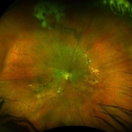
Fundus photograph showing bony spicules in a case of Retinitis Pigmentosa.
Condition/keywords: Bony spicules, Retinitis Pigmentosa (RP)
-
 Retinitis Pigmentosa (RP)
Sep 14 2023 by Ben Serar
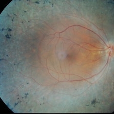
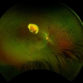
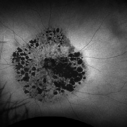
Fundus photograph of the RE showing bony spicules in the mid-periphery, with waxy disc pallor and arteriolar attenuation in a case of retinitis pigmentosa.
Condition/keywords: Retinitis Pigmentosa (RP)
-
 Retinitis Pigmentosa (RP)
Sep 12 2023 by Ben Serar
Fundus photograph showing bony spicules and arteriolar attenuation in a case of Retinitis Pigmentosa.
Condition/keywords: arteriolar attenuation, Bony spicules, Retinitis Pigmentosa (RP)
-
 A Feast for Crows , Retinitis pigmentosa
Sep 22 2022 by wang xiaomei
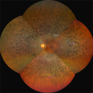
Fundus photograph of an 55-year-old man with Retinitis Pigmentosa, There is increasing loss of pigment from the pigment epithelium with intraretinal clumping of melanin, appearing most often as coarse clumps in a "bone spicule" configuration, arteriolar narrowing
Photographer: Man, Li, Bao Ji Ophthalmic Hospital
Imaging device: ZEISS CLARUS 500
Condition/keywords: retinitis pigmentosa (RP) dystrophy
-
 Bone Corpuscle Pigments
Sep 11 2014 by Mehul A Shah
A 42-year-old female presented with gradual reduction in vision.
Photographer: Drashti Netralaya,Dahod
Imaging device: FF 450
Condition/keywords: retinitis pigmentosa (RP) dystrophy
-
 RETINITIS PIGMENTOSA
Oct 15 2022 by Akansha Sharma
COLOUR FUNDUS PHOTOGRAPH OF A 30 YEAR OLD MALE WITH RETINITIS PIGMENTOSA
Photographer: Dr. Akansha Sharma-Retina Foundation, Ahmedabad
Condition/keywords: retinitis pigmentosa (RP) dystrophy, RP variant
-
 RETINITIS PIGMENTOSA
Oct 15 2022 by Akansha Sharma
COLOUR FUNDUS PHOTOGRAPH OF A 30 YEAR OLD MALE WITH RETINITIS PIGMENTOSA
Photographer: Dr. Akansha Sharma-Retina Foundation, Ahmedabad
Condition/keywords: retinitis pigmentosa (RP) dystrophy, RP variant
-
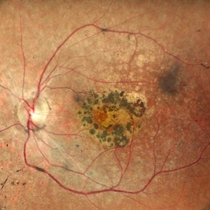
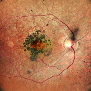
 Retinitis Pigmentosa With Macular Involvment
Sep 11 2014 by Mehul A Shah
A 35-year-old female presented with gradual reduction in vision.
Photographer: Drashti Netralaya,Dahod
Imaging device: FF 450
Condition/keywords: retinitis pigmentosa (RP) dystrophy
-
 RP Retisert
Feb 2 2017 by Jeffrey L. Olson, MD
20-year-old patient with retinitis pigmentosa and pars planitis, recently s/p Retisert implant
Photographer: William Yates, University of Colorado Eye Center
Imaging device: Optos California
Condition/keywords: retinitis pigmentosa (RP) dystrophy
-
 RP with VPT
Jul 29 2023 by Mohammadkarim Johari
25 years old boy, with past history of RP, recently he came with decreased vision in Rt eye.
Photographer: Mohammadkarim Johari, Shiraz university of medical science
Condition/keywords: retinitis pigmentosa (RP) dystrophy, vascular anomaly, vasoproliferative retinopathy, vasoproliferative vitreoretinopathy
-
 Unilateral Retinitis Pigmentosa
May 1 2014 by Raj K. Maturi, MD
53-year-old woman with significant salt and pepper retinopathy OS.
Photographer: Tom Steele Midwesteye Institute 200 w 103rd st Indianapolis, Indiana
Condition/keywords: retinitis pigmentosa (RP) dystrophy
-
 Unilateral Retinitis Pigmentosa
May 1 2014 by Raj K. Maturi, MD
53-year-old woman with significant salt and pepper retinopathy OS.
Photographer: Tom Steele, Midwest Eye Institute
Condition/keywords: retinitis pigmentosa (RP) dystrophy
-
 Advanced Retinitis Pigmentosa
Mar 14 2017 by Hamid Ahmadieh, MD
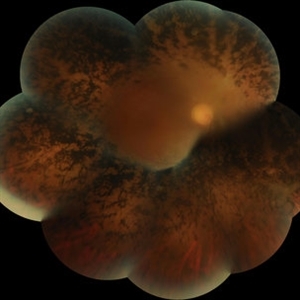
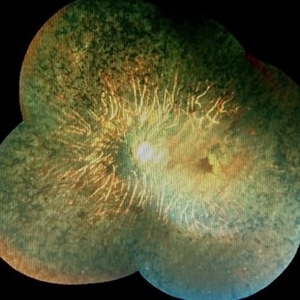
Merged color fundus photograph of the right eye of a patient with advanced retinitis pigmentosa sparing the posterior pole.
Photographer: Soodabeh Fouladin, Negah Eye Center, Tehran, Iran
Condition/keywords: color fundus photograph, retinitis pigmentosa (RP) dystrophy
-
 Inflammation
May 15 2013 by Howard Schatz, MD
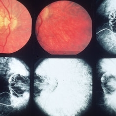
30-year-old white female, 20/30 periphery and posterior RP dystrophy.
Condition/keywords: inflammation, retinitis pigmentosa (RP) dystrophy
-
 Inflammation
May 15 2013 by Howard Schatz, MD
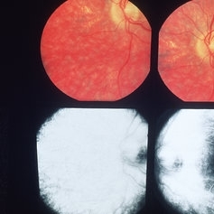
30-year-old white female, 20/50 periphery and posterior RP dystrophy.
Condition/keywords: inflammation, retinitis pigmentosa (RP) dystrophy
-
 Mucopolysaccharidosis Type III
Apr 21 2023 by Matthew Dombrow, MD
Fundus photograph and autofluorescence of a 49 year old male with mucopolysaccharidosis type III (Sanfilippo syndrome)
Photographer: Cori Sturtevant, Connecticut Retina Consultants, Hamden, Connecticut
Condition/keywords: mucopolysaccharidoses, retinitis pigmentosa (RP) dystrophy
-
 Mucopolysaccharidosis Type III (Sanfilippo syndrome)
Apr 21 2023 by Matthew Dombrow, MD
Fundus photograph and autofluorescence of a 49 year old male with mucopolysaccharidosis type III (Sanfilippo syndrome)
Photographer: Cori Sturtevant, Connecticut Retina Consultants, Hamden, Connecticut
Condition/keywords: mucopolysaccharidoses, retinitis pigmentosa (RP) dystrophy
-
 Retinitis Pigmentosa
Apr 30 2015 by Mitzy E Torres Soriano, MD
Fundus of patient with retinitis pigments, bone spicule-shaped pigment deposits are present with retinal atrophy, while the macula is preserved . Retinal vessels are attenuated.
Photographer: Mitzy E. Torres Soriano, MD; Centro medico Cagua-Estado Aragua. Venezuela
Imaging device: TRC-NW8
Condition/keywords: pigmentary retinal dystrophy, retinal dystrophy, retinitis pigmentosa, retinitis pigmentosa (RP) dystrophy
-
 Retinitis Pigmentosa
Aug 25 2015 by René Hernán Parada Vásquez
Fundus photograph of both eyes of a 38-year-old female with retinitis pigmentosa, bone spicule-shaped pigment deposits are present in the mid periphery, and macula with a peripheral ring of depigmentation.
Photographer: Parada René, ESO, Guatemala.
Imaging device: Canon CR-2
Condition/keywords: bilateral pigmentary retinopathy, retinitis pigmentosa, retinitis pigmentosa (RP) dystrophy
-
 Retinitis pigmentosa
Feb 26 2020 by Manuel Ángel Alcántara Delgado, MD
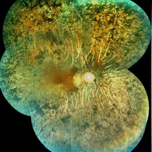
Merged color fundus photograph of a 68-year-old woman with advanced retinitis pigmentosa. It is appreciated bone spicule-shaped pigment deposits, optic disc pallor, retinal atrophy and attenuated retinal vessels.
Photographer: Manuel Ángel Alcántara Delgado
Condition/keywords: choroidal circulation, optic disc pallor, pericentral retinitis pigmentosa, retina, retinitis pigmentosa, retinitis pigmentosa (RP) dystrophy, sector retinitis pigmentosa
-
 Retinitis Pigmentosa
Feb 26 2020 by Manuel Ángel Alcántara Delgado, MD
Merged color fundus photograph of a 68-year-old woman with advanced retinitis pigmentosa. It is appreciated bone spicule-shaped pigment deposits, optic disc pallor, retinal atrophy, attenuated retinal vessels and surface wrinkling retinopathy.
Photographer: Manuel Ángel Alcántara Delgado
Condition/keywords: chorioretinal atrophy, choroidal circulation, optic disc pallor, pericentral retinitis pigmentosa, retina, retinitis pigmentosa, retinitis pigmentosa (RP) dystrophy, sector retinitis pigmentosa
 Loading…