Search results (16 results)
-
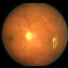 Bidding Adieu to Attachments: Weiss Ring
Jan 7 2022 by Gayathri Mohan
Colour fundus photograph showing a Weiss ring following PVD.
Photographer: Dr. GAYATHRI MOHAN
Imaging device: Canon
Condition/keywords: PVD induction, Weiss ring
-
 Detached NVE During PVD induction
Apr 27 2018 by Michael J. Koss, MD, PhD, MBA
A 73-year-old woman with macular pucker underwent a pars plana vitrectomy with membrane peeling. Additionally the patient suffers from diabetic retinopathy after being diagnosed with type 2 diabetes mellitus sixteen years ago. Prior to the procedure she was treated with a series of intravitreal Bevacizumab-injections due to diabetic macular edema. There was no history of a proliferative DRP. During the vitrectomy a branch of an obliterated NVE spontaneously detached and floated freely in the vitreous. The 3D shot was captured via Alcon’s NGENUITY® 3D Visualization System in form of photograph and video providing an outstandingly detailed image of the branched NVE.
Photographer: Michael Koss, Augenzentrum Nymphenburger Hoefe
Imaging device: Alcon’s NGENUITY® 3D Visualization System
Condition/keywords: diabetes, diabetic retinopathy, neovascularization elsewhere (NVE), pars plana vitrectomy (PPV), PVD induction
-
 Epiretinal Membrane
Feb 2 2022 by Manish Nagpal, MD, FRCS (UK), FASRS
Intraoperative photo of a epiretinal membrane, glistening reflex noted. Prior to this capture, PVD induction has been done, which has left a small splinter hemorrhage around the disc attachment of hyaloid.
Photographer: Manish Nagpal, Retina Foundation, Ahmedabad, India
Imaging device: Sony PMW -10 MD surgical camera
Condition/keywords: epiretinal membrane formation, ERM, ILM flap, PVD induction
-
 Intraoperative Triamcinolone Staining to Visualize Hyaloid
Jan 10 2022 by Manish Nagpal, MD, FRCS (UK), FASRS
Intraoperative image of triamcinolone being injected in order to stain the hyaloid for facilitating PVD induction.
Photographer: Manish Nagpal, Retina Foundation, Ahmedabad, India
Imaging device: Sony PMW -10 MD surgical camera
Condition/keywords: hyaloid, PVD induction, triamcinolone
-
 Posterior Vitreous Detachment
Mar 21 2019 by Michael Politis, MD
Intra-op image of a PVD induction using Kenalog in a retinal detachment after retained nuclear material
Photographer: Michael Politis MD, McGill University, Montreal, Canada
Imaging device: Karl Stroze
Condition/keywords: kenalog, PVD induction
-
 Posterior Vitreous Detachment
Jan 31 2025 by Thirumalesh Mochi Basavaraj, MD
Intraoperative view of Triamcinolone-assisted posterior vitreous detachment.
Photographer: Thirumalesh Mochi Basavaraj
Condition/keywords: PVD induction, triamcinolone
-
 PVD Induction
Feb 2 2022 by Manish Nagpal, MD, FRCS (UK), FASRS
Intraoperative photo of PVD induction being carried out. Hyaloid has been stained using triamcinolone dye and PVD induction is carried out using high suction on the cutter and engaging the stained hyaloid.
Photographer: Manish Nagpal, Retina Foundation, Ahmedabad, India
Imaging device: Sony PMW -10 MD surgical camera
Condition/keywords: Hyaloid staining, PVD induction, triamcinolone
-
 PVD Induction in Progress
Jan 10 2022 by Manish Nagpal, MD, FRCS (UK), FASRS
Intraoperative image of PVD induction with triamcinolone staining clearly showing the Weiss ring, which is being lifted with suction from the cutter.
Photographer: Manish Nagpal, Retina Foundation, Ahmedabad, india
Imaging device: Sony PMW -10 MD surgical camera
Condition/keywords: PVD induction, triamcinolone, Weiss ring
-
 PVD induction with IVTA staining
Nov 1 2022 by Shobhit Chawla, M.S.
This is an intraoperative photograph of Pvd induction showing both the macular and disc attatchments stained with triamcinolone.
Photographer: Shobhit Chawla
Condition/keywords: PVD induction, triamcinolone
-
 Triamcinolone Acetonide stained PVD induction by cutter
Apr 11 2014 by Subhendu Kumar Boral, MBBS, MD(AIIMS), DNB, FASRS (USA)
Intra operative step of PVD induction in a case of diabetic epiretinal membrane in Left Eye in a 68 years old gentleman
Photographer: Subhendu Kumar Boral
Condition/keywords: PVD induction
-
PVD induction in a retinal detachment
Oct 24 2022 by Manish Nagpal, MD, FRCS (UK), FASRS
This video highlights the PVD induction technique in a case of retinal detachment with mobile retina, triamcinolone staining allows ease of visualizing the pvd attachment which is gradually removed from the retinal attachment using suction.
Photographer: Manish Nagpal
Condition/keywords: posterior hyaloid, PVD, triamcinolone, video, vitrectomy
-
Macular Hole Surgery: Inverse Flaps
Jan 31 2025 by Thirumalesh Mochi Basavaraj, MD
This video demonstrates, PVD induction , followed by ILM peeling in multiple flower petal flap technique.
Condition/keywords: ILM flaps, ILM peel, induction
-
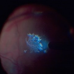
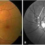 Peri-papillary Vascular Loop
Jun 2 2020 by Dhaivat Shah
Peri-papillary vascular loops (PVL) are rare congenital vascular malformations, which are usually detected as accidental finding during routine fundus examination. They can often be confused with tributary vein occlusion or racemose hemangioma. Although benign and asymptomatic, they can be rarely associated with vitreous hemorrhage and arterial occlusion. We herein present a case of a 60-year-old hypertensive male, who was diagnosed elsewhere to have a tributary vein occlusion and was referred to us. FFA was advised to rule out neovascularization, surrounding capillary non perfusion and mass lesion (hemangioma). On FFA, the arterial loop showed a slightly delayed filling (3-5 seconds) as compared to the other arterial vessels and the original vessel appeared to be a branch arising from central retinal artery. The choroidal filling was delayed in the area supplied by the loop. A cilioretinal artery was also noted. The patient was diagnosed to have a Peri-papillary vascular arterial loop (PVL), likely to be congenital in origin. The patient was reassured and was advised yearly follow up. These loops are usually accidental findings discovered during routine fundus examination. Since these vessels are looped and tortuous, they exhibit a slower and laminar blood flow, which make them more prone for arterial occlusions. The vitreous in this area tends to be adherently attached, so during PVD induction, it is likely to cause a tear and hemorrhage leading to vitreous hemorrhage. Until and unless there is a break, this hemorrhage tends to resolve on its own and does not warrant treatment. If there is an evident break, it can be dealt with laser barrage.
Photographer: Choithram Netralaya
Condition/keywords: congenital prepapillary vascular loop
-
 TA Stained Posterior Hyaloid Face
Apr 11 2014 by Subhendu Kumar Boral, MBBS, MD(AIIMS), DNB, FASRS (USA)
Intraoperative step of posterior hyaloid face staining by triamcinolone acetonide particles during PVD induction in a case of diabetic epiretinal membrane left eye in a 68-year-old gentleman.
Photographer: Subhendu Kumar Boral
Condition/keywords: hyaloid
-
Vitrectomy for epiretinal membrane removal
Nov 30 2022 by Manish Nagpal, MD, FRCS (UK), FASRS
Vitrectomy is carried out for epiretinal membrane removal. After doing core vitrectomy and pvd induction a 25 gauge forceps is used to pinch and peel the macular pucker. After finding a edge the forceps is gradually moved tangentially over the retinal surface to remove the membrane.
Photographer: Manish Nagpal
Condition/keywords: epiretinal membrane, forceps, macular pucker, peeling, video, vitrectomy
-
Vitrectomy for Macular Hole
Jan 13 2023 by Manish Nagpal, MD, FRCS (UK), FASRS
This is a case of Macular hole for which vitrectomy is being done. After doing core vitrectomy triamcinolone dye is injected to stain the hyaloid. High aspiration is used on cutter to engage the hyaloid and gradually pull it anteriorly. PVD induction is carried out. After this brilliant blue dye is injected to stain the internal limiting membrane. ILM is peeled using a 25 gauge forceps in a tangential manner. After this i use a instrument called the massager which we have developed to gently and atraumatically massage concentrically the edges of the hole. This releases the subtle contaction on the edges of the hole and relaxes the margins. After this air fluid exchange is carried out followed by low vacuum aspiration over the hole. The hole approximates itself gradually as the aspiration dries up the edges.
Condition/keywords: forceps, hyaloid, ILM, macular hole, peeling, staining, video, vitrectomy
 Loading…