Search results (521 results)
-
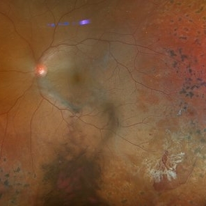
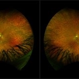 "Mud-Splatter" of Posterior Pole and Peripheral Radial Streaks in a Carrier of Ocular Albinism
Jan 22 2019 by John S. King, MD
14-year-old healthy white female with family history of ocular albinism was seen by Dr. Hruby for a second opinion. Father and some of his brothers were positive for a history of ocular albinism. Va cc 20/30 J1+ OU; no nystagmus; no TIDs; no foveal hypoplasia. A "mud-spatter" appearance to the posterior pole was present, along with peripheral alternating streaks (photo). Dr. Hruby agreed that this was most likely a carrier of Ocular Albinism Type-1 (XR; GPR143 mutation), and possible genetic testing/counselling was discussed.
Photographer: Gretchen Harper
Imaging device: Optos California
Condition/keywords: Nettleship-Falls ocular albinism, ocular albinism
-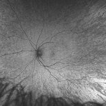
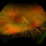
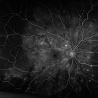
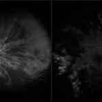
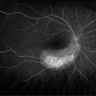
 "Mud-Splatter" of Posterior Pole and Peripheral Radial Streaks in a Carrier of Ocular Albinism
Jan 22 2019 by John S. King, MD
14-year-old healthy white female with family history of ocular albinism was seen by Dr. Hruby for a second opinion. Father and some of his brothers were positive for a history of ocular albinism. Va cc 20/30 J1+ OU; no nystagmus; no TIDs; no foveal hypoplasia. A "mud-spatter" appearance to the posterior pole was present, along with peripheral alternating streaks, which are very prominent in this late phase FA of the right eye. Dr. Hruby agreed that this was most likely a carrier of Ocular Albinism Type-1 (XR; GPR143 mutation), and possible genetic testing/counselling was discussed.
Photographer: Gretchen Harper
Imaging device: Optos California
Condition/keywords: Nettleship-Falls ocular albinism, ocular albinism
-
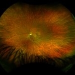
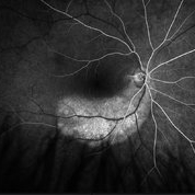
 "Mud-Splatter" of Posterior Pole and Peripheral Radial Streaks in a Carrier of Ocular Albinism
Jan 22 2019 by John S. King, MD
14-year-old healthy white female with family history of ocular albinism was seen by Dr. Hruby for a second opinion. Father and some of his brothers were positive for a history of ocular albinism. Va cc 20/30 J1+ OU; no nystagmus; no TIDs; no foveal hypoplasia. A "mud-spatter" appearance to the posterior pole was present, along with peripheral alternating streaks that are very prominent on this late phase FA OS. Dr. Hruby agreed that this was most likely a carrier of Ocular Albinism Type-1 (XR; GPR143 mutation), and possible genetic testing/counselling was discussed.
Photographer: Gretchen Harper
Imaging device: Optos California
Condition/keywords: Nettleship-Falls ocular albinism, ocular albinism
-
 "Mud-Splatter" of Posterior Pole and Peripheral Radial Streaks in a Carrier of Ocular Albinism
Jan 22 2019 by John S. King, MD
14-year-old healthy white female with family history of ocular albinism was seen by Dr. Hruby for a second opinion. Father and some of his brothers were positive for a history of ocular albinism. Va cc 20/30 J1+ OU; no nystagmus; no TIDs; no foveal hypoplasia. A "mud-spatter" appearance to the posterior pole was present, along with peripheral alternating streaks (photo). Dr. Hruby agreed that this was most likely a carrier of Ocular Albinism Type-1 (XR; GPR143 mutation), and possible genetic testing/counselling was discussed.
Photographer: Gretchen Harper
Imaging device: Optos California
Condition/keywords: Nettleship-Falls ocular albinism, ocular albinism
-
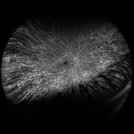
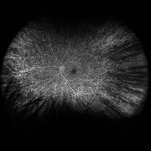
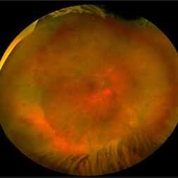
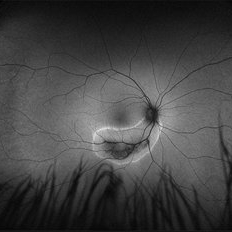
 "Mud-Splatter" of Posterior Pole and Peripheral Radial Streaks in a Carrier of Ocular Albinism
Jan 22 2019 by John S. King, MD
14-year-old healthy white female with family history of ocular albinism was seen by Dr. Hruby for a second opinion. Father and some of his brothers were positive for a history of ocular albinism. Va cc 20/30 J1+ OU; no nystagmus; no TIDs; no foveal hypoplasia. A "mud-spatter" appearance to the posterior pole was present, along with peripheral alternating streaks. Hypoautofluorescent areas correspond to hyperpigmented areas of retinal pigment epithelium, and vice versa (see photo). Dr. Hruby agreed that this was most likely a carrier of Ocular Albinism Type-1 (XR; GPR143 mutation), and possible genetic testing/counselling was discussed.
Photographer: Gretchen Harper
Imaging device: Optos California
Condition/keywords: Nettleship-Falls ocular albinism, ocular albinism
-
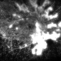
 "Starry Sky" Fundus in Vogt-Koyanaki-Harada Syndrome
Jan 10 2018 by Peter H. Tang, MD, PhD
Fluorescein angiography imaging of a 27-year-old male with acute inflammation as part of Vogt-Koyanagi-Harada Syndrome.
Imaging device: Optos California
Condition/keywords: chorioretinal inflammations, retina, uveitis, Vogt-Koyanagi-Harada
-
 Active neovascularization in Proliferative Diabetic Retinopathy
Jan 10 2018 by Peter H. Tang, MD, PhD
Fluorescein angiography image from a 46-year-old woman with uncontrolled proliferative diabetic retinopathy shows extensive dye leakage from active neovascularization.
Imaging device: Optos California
Condition/keywords: diabetes, diabetic retinopathy, fluorescein leakage, neovascularization elsewhere (NVE), neovascularization of the disc (NVD), pan-retinal photocoagulation (PRP), proliferative diabetic retinopathy (PDR)
-
 Actively Bleeding NVE
Apr 1 2025 by Jordyn Beckman
47 year old woman presented with actively bleeding NVE temporally on exam with complaints of foggy vision and floaters.
Photographer: Jordyn Beckman, Retina Consultants of Carolina, P.A.
Imaging device: Optos California
Condition/keywords: active bleeding, Elevated retinal neovascularization, vitreous hemorrhage
-
 Acute Central Retinal Artery Occlusion
Jul 27 2022 by Becca Harris
Ultra widefield FA/ICG of a 24 year old female with an acute central retinal artery occlusion affecting the right eye. Patient presented with extreme headaches following DAVF surgery the previous day. Patient has Factor VIII deficiency and had a cerebral venous thrombosis 9 years ago and lost vision in the right eye at that time. Patient has history of optic sheath fenestration OU and craniotomy. On initial evaluation, she had a CRAO as well as diffuse choroidal nonperfusion noted on optos FA. Suspect nonperfusion to third and sixth nerve leading to palsy. Occlusion of vasculature in the setting of recent endovascular embolization of fistulas in the CNS. Discussed diagnosis and poor prognosis with parents and patient. Patient had no light perception at the time of her initial appointment.
Photographer: Becca Harris
Imaging device: Optos California
Condition/keywords: Choroidal non-perfusion, fluorescein angiogram (FA), indocyanine green (ICG) angiography, non-perfusion, Optos, Right Eye, ultra-wide field imaging
-
 Acute Posterior Multifocal Placoid Pigment Epitheliopathy
Feb 20 2024 by Soobien Lee
Optos fundus autofluorescence photograph of a 20-year-old caucasian female with viral prodrome and vision loss OS>OD secondary to Acute Posterior Multifocal Placoid Pigment Epitheliopathy (APPME). Imaging of her left eye shows hypoautofluorescent areas corresponding to multiple bilateral placoid lesions at the level of RPE and choroid throughout the posterior pole.
Photographer: Ashley Metzger, Elman Retina Group
Imaging device: Optos Ultra-Widefield Autoflurescence Imaging
Condition/keywords: acute posterior multifocal placoid pigment epitheliopathy (APMPPE), autofluorescence imaging, bacilliary layer detachment, Optos, OPTOS CALIFORNIA, uveitis, white dot syndrome
-
 Acute Retinal Necrosis (resolved)
Aug 21 2025 by Virginia Gebhart
31 year old female with resolved Acute Retinal Necrosis. Pt was positive for HSV2 in 2019, received intravitreal Ganciclovir x3. Both eyes remain stable with no active inflammation, retinitis remains inactive with pigmented edges. Pt continues taking Valtrex qday for maintenance. BCVA 20/40
Photographer: Virginia Gebhart, Retina Consultants of Carolina
Imaging device: Optos California
Condition/keywords: acute retinal necrosis, retinitis
-
 Acute Retinal Necrosis secondary to Herpes Zoster Ophthalmicus
Jan 9 2018 by Olivia Rainey
Ultra-wide field Optos pseudocolor montage of an 40-year-old female presenting with acute retinal necrosis secondary to herpes zoster ophthalmicus affecting her right eye.
Photographer: Olivia Rainey
Imaging device: Optos California
Condition/keywords: acute retinal necrosis, color fundus photograph, Herpes zoster, montage, Optos, ultra-wide field imaging
-
 Acute Zonal Occult Outer Retinopathy (AZOOR)
Jan 19 2022 by James B. Soque, CRA, OCT-C, COA, FOPS
Acute Zonal Occult Outer Retinopathy, OD. Color fundus, Ultra-wide field photograph. 46-year-old white male, VA CC 10/16, 20/12.5, has had recurrent vasculitis for 11 years. No treatment.
Photographer: James Soque, CRA, OCT-C, COA, FOPS, Island Retina, Shirley, NY
Imaging device: Optos California
Condition/keywords: acute zonal occult outer retinopathy (AZOOR), color wide field, optos, ultra-wide field imaging
-
 Acute Zonal Occult Outer Retinopathy (AZOOR) FA, Fluorescein Angiography, Peripheral Vasculitis
Jan 19 2022 by James B. Soque, CRA, OCT-C, COA, FOPS
Acute Zonal Occult Outer Retinopathy (AZOOR). Peripheral Vasculitis OD. Fluorescein angiography showing vasculitis in the far right periphery 8-10 o'clock. 46-year-old white male, VA CC 20/16, 20/12.5, has had recurrent vasculitis for 11 years. No treatment.
Photographer: James Soque, CRA, OCT-C, COA, FOPS, Island Retina, Shirley, NY
Imaging device: Optos California
Condition/keywords: acute zonal occult outer retinopathy (AZOOR), FA early phase, fluorescein angiogram (FA), Peripheral Vasculitis, ultra-wide field imaging
-
 Acute Zonal Occult Outer Retinopathy (AZOOR) FA, Ultra Wide-Field Fluorescein Angiogram Early
Jan 19 2022 by James B. Soque, CRA, OCT-C, COA, FOPS
Acute Zonal Occult Outer Retinopathy, FA, Fluorescein Angiography, OD. 46-year-old white male, VA CC 10/16, 20/12.5, has had recurrent vasculitis for 11 years. No treatment.
Photographer: James Soque, CRA, OCT-C, COA, FOPS, Island Retina, Shirley, NY
Imaging device: Optos California
Condition/keywords: acute zonal occult outer retinopathy (AZOOR), FA EARLY, fluorescein angiogram (FA), ultra-wide field imaging
-
 Acute Zonal Occult Outer Retinopathy, (AZOOR) FAF, Fundus Autofluorescence
Jan 19 2022 by James B. Soque, CRA, OCT-C, COA, FOPS
Acute Zonal Occult Outer Retinopathy, FAF, Fundus Auto Fluorescence, OD. 46-year-old white male, VA CC 10/16, 20/12.5, has had recurrent vasculitis for 11 years. No treatment.
Photographer: James Soque, CRA, OCT-C, COA, FOPS, Island Retina, Shirley, NY
Imaging device: Optos California
Condition/keywords: acute zonal occult outer retinopathy (AZOOR), fundus autofluorescence (FAF), ultra-wide field imaging
-
 Adult Onset Coats' Disease
Jul 5 2024 by Zach Seim
FA/ICG of a 64 year old female with Adult Onset Coats' Disease. VA DCC CF@3 feet upon presentation. Therapy options discussed extensively.
Photographer: Zach Seim
Imaging device: Optos California
Condition/keywords: Coats' disease, FA, indocyanine green (ICG) angiography, Optos, OPTOS CALIFORNIA
-
 Asteroid Hyalosis
Jul 5 2024 by Zach Seim
Optos fundus photo of an 88 year old male with Asteroid Hyalosis OS. VA Dsc 20/30-2 upon presentation.
Photographer: Zach Seim
Imaging device: Optos California
Condition/keywords: ASTEROID, asteroid hyalosis, optos, OPTOS CALIFORNIA
-
 Asteroid Hyalosis
Oct 8 2025 by Virginia Gebhart
49 year old female with asteroid hyalosis. Pt only notices when in bright light.
Photographer: Virginia Gebhart, Retina Consultants of Carolina
Imaging device: Optos California
Condition/keywords: asteroid hyalosis
-
 Asteroid Hyalosis in Retinitis Pigmentosa
Dec 9 2024 by Mauricio Bayram-Suverza, MD
A 54 year-old male patient presented with asteroid hyalosis. Retinal examination revealed the presence of bone spicules, primarily located in the mid-periphery. Genetic testing identified a pathogenic variant in the RHO gene.
Photographer: Mauricio Bayram-Suverza, Casey Eye Institute, OHSU.
Imaging device: Optos California
Condition/keywords: Asteroid hyalosis, retinal dystrophy, Retinitis Pigmentosa, vitreous
-
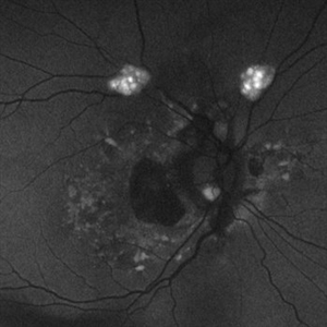
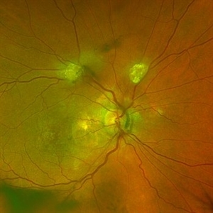 Astrocytic Hamartoma
Feb 27 2025 by Daniel Davis, OCT-C
Color fundus photo of 55-year-old female with Astrocytic Hamartoma in association with tuberous sclerosis. No treatment options available, benign. Other findings include; Posterior Vitreous Detachment, Vitreous Hemorrhage, Hereditary Retinal Dystrophy, Vitreous Opacities, Hypertensive Retinopathy.
Photographer: Daniel Davis, OCT-C
Imaging device: Optos California
Condition/keywords: color fundus photograph
-
 Astrocytic Hamartoma
Feb 27 2025 by Daniel Davis, OCT-C
Fundus autofluorescence photo of 55-year-old female with astrocytic hamartoma in association with tuberous sclerosis. No treatment options available, benign. Other findings include; Posterior Vitreous Detachment, Vitreous Hemorrhage, Hereditary Retinal Dystrophy, Vitreous Opacities, Hypertensive Retinopathy.
Photographer: Daniel Davis, OCT-C
Imaging device: Optos California
Condition/keywords: astrocytic hamartoma, fundus autofluorescence (FAF)
-
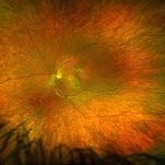
 BB Gun Accident
Dec 13 2017 by Jason Griffith
8-year-old male patient presented with history of metallic extraocular foreign body OD x 3 weeks; patient voiced history of being struck by BB approximately 3 weeks prior to exam; pt. was taken to OR for removal of metallic intraocular foreign body.
Photographer: Jason Griffith
Imaging device: Optos California
Condition/keywords: chorioretinitis sclopetaria
 Loading…