Search results (13 results)
-
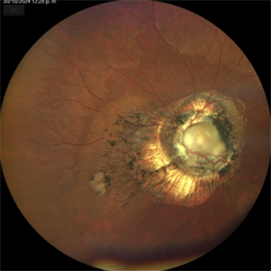
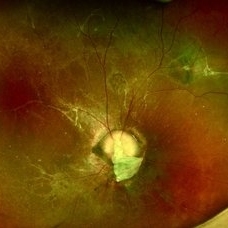
 Coexistent Morning Glory Syndrome and Persistent Hyperplastic Primary Vitreous
Jul 15 2024 by Arthi Mohankumar , MS,MRCS ED, FICO,FAICO
Anterior segment and fundus photograph of a 9-year-old female revealing PHPC changes (Images A,B,C) and morning glory syndrome (image D).
Photographer: Arthi Mohankumar
Condition/keywords: Morning Glory Anomaly, Morning Glory Syndrome, persistent hyperplastic primary vitreous (PHPV)
-
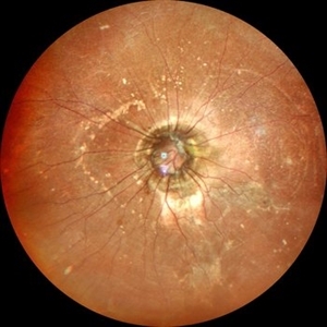
 Morning Glory Anomaly
Dec 31 2016 by Linda A Cernichiaro- Espinosa, MD
Morning Glory Anomaly
Photographer: Paulina Tolosa T
Imaging device: Optos
Condition/keywords: Morning Glory Syndrome, vitreoretinal degeneration
-
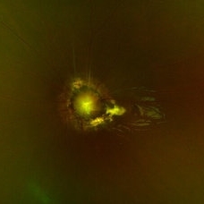
 Morning Glory Anomaly
Mar 24 2022 by Elite Bor-Shavit, MD
Disc photo of a 28-years-old male with Morning Glory Anomaly of his right optic nerve observed over time.
Condition/keywords: Morning Glory Anomaly, optic disc
-
 Morning Glory Anomaly
Sep 20 2021 by Luis Daniel Gutierrez, MD
Ultra-wide field color clinical photograph of the left eye with Morning Glory anomaly of the optic nerve and subretinal fluid in the macular area.
Photographer: Luis Daniel Gutierrez García, Hospital Fundación Nuestra Señora de la Luz, Ciudad de México.
Imaging device: Optos
Condition/keywords: congenital malformation of the optic nerve, Morning Glory Anomaly
-
 Morning Glory Anomaly
Jan 5 2025 by César Adrián Gómez Valdivia, MD
Morning Glory Anomaly found in a 10 year-old male patient with poor visual acuity and strabismus. Findings were unilateral.
Photographer: @eyemissu2
Imaging device: TOPCON TRC-50DX
Condition/keywords: Morning Glory Anomaly, Morning Glory Syndrome
-
 Morning Glory Anomaly with Macular Atrophy
Oct 30 2024 by Luis Guillermo Anaya Sánchez, Ophthalmology
47 year old female patient, presents to evaluate low vision since birth. Fundus photography shows an enlarged disc, with radial vasculature disposition, and glial tissue; corresponding with a Morning Glory Anomaly, with macular atrophy.
Photographer: Luis Guillermo Anaya MD
Imaging device: Zeiss Clarus 700
Condition/keywords: Morning Glory Anomaly
-
 Morning Glory Anomaly With Retinal Detachment Managed With Amniotic Membrane Graft
Oct 15 2024 by Hemanth Murthy, MBBS, MD, FASRS
10 year-old boy presented with noticed blurring of vision. He had total retinal detachment with complicated cataract. He underwent lensectomy with 240 band and vitrectomy with silicone oil. The retina failed to settle due to minute breaks in the inferior part of the disc. Repeat surgery with AMG was done to cover the inferior part of disc. The retina settled under silicone oil. Silicone oil was removed and he is presently undergoing amblyopia treatment. Vision is 2/60 with +14.5 diopter lens.
Photographer: Mr Veda Vyas
Condition/keywords: amniotic membrane graft, Morning Glory Anomaly
-
 Morning Glory Disc
Jun 26 2022 by Vaidehi Sathaye
Fundus photograph of a 5 yr old male child with Morning Glory Disc with Subretinal Fluid
Photographer: Dr. Vaidehi Sathaye
Imaging device: Mirante
Condition/keywords: Morning Glory Anomaly
-
 Morning Glory Disc Anomaly
Dec 15 2023 by Brandon I Fram, MD, BS
Fundus photograph of an isolated morning glory disc anomaly
Condition/keywords: Morning Glory Anomaly
-
 Morning Glory Disc Anomaly
Feb 12 2024 by NIDHI PANWAR, MD FNB FICO
Fundus photograph of 43 year old male, hypertensive on medication, came for routine check up, and has been diagnosed to have poor vision left eye since childhood, denies any history of trauma. Vision left eye 6/18, Anterior segment normal, Fundus left eye shows excavated ,funnel-shaped optic nerve head, with central tuft of glial tissue obscuring the cup . The retinal vessels were seen emanating from the edge of disc in radial manner. In addition, the sectoral nasal retina shows localized area of hyperpigmented bony spicules like lesions. However, no history of nyctalopia or any other neurological disorder could be obtained.
Photographer: Nidhi Panwar, NMC Royal hospital, Sharjah , UAE
Imaging device: OPTOMAP
Condition/keywords: Morning Glory Anomaly, optic disc excavation
-
 Morning glory disc anomaly-associated maculopathy: fibroglial tissue with a Mac-Off serous retinal detachment.
Jun 26 2024 by JULIAN VILLARREAL, MD
19 year old with a Morning glory disc anomaly-associated maculopathy: fibroglial tissue with a Mac-Off serous retinal detachment.
Photographer: Julián Villarreal MD
Imaging device: Mirante
Condition/keywords: fibroglial tissue, Morning Glory Anomaly, retinal detachment of the macula
-
 Morning glory optic disc anomaly with retinal detachment
Sep 13 2022 by Min Kim, MD, PhD, MBA, FASRS
Fundus examination of this 5 year-old male shows large funneled optic nerve with conical excavation of the dysplastic optic disc. 360° macula-involving retinal detachment was observed. The best corrected visual acuity of the right eye was counting fingers 10cm.
Photographer: Min Kim, M.D.-Ph.D.-M.B.A. Gangnam Severance Hospital Yonsei University College of Medicine, Department of Ophthalmology
Imaging device: Optos Silverstone P200TxE
Condition/keywords: Morning Glory Anomaly, Morning Glory Syndrome
-
 Rhegmatogenous retinal detachment with dislocated IOL in a Morning Glory anomaly
Jul 27 2023 by Gustavo Aguirre-Suarez
Fundus photograph of a 13-year-old male with a history of congenital cataract surgery in his right eye in 2019. The patient presents with sudden visual loss. Upon examination, a dislocated IOL is observed in the posterior segment, accompanied by a rhegmatogenous retinal detachment featuring peripheral retinal tears and horseshoe breaks. Additionally, a morning glory disc anomaly is also present in this patient.
Photographer: Gustavo Aguirre-Suarez
Imaging device: Mirante, NIDEK
Condition/keywords: dislocated posterior chamber intraocular lens (PCIOL), Morning Glory Anomaly, rhegmatogenous retinal detachment
 Loading…