Search results (7 results)
-
 Malignant Melanoma
Sep 11 2018 by Olivia Rainey
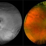
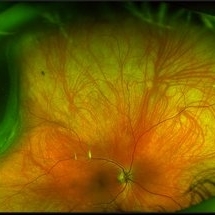
Ultra-wide field autofluorescence and pseudocolor montage of a 57-year-old male s/p I-125 brachytherapy for malignant melanoma affecting his right eye. The patient’s radiation retinopathy has resulted in retinal vascular occlusive disease and optic nerve edema.
Photographer: Olivia Rainey
Imaging device: Optos
Condition/keywords: branch retinal vein occlusion (BRVO), fundus autofluorescence (FAF), I-125 brachytherapy, malignant melanoma, montage, Optos, pseudocolor, radiation retinopathy, ultra-wide field imaging
-
 Radiation Retinopathy Post Brachytherapy Plaque Treating Choroidal Melanoma
May 26 2020 by Sophia El Hamichi, MD
A 71-year-old female treated with plaque brachytherapy for choroidal melanoma in her left eye 10 years ago. She subsequently developed radiation retinopathy OS for which she was regularly receiving intravitreal injections of bevacizumab. The result is a stable visual acuity at 20/30. The patient continues to be regularly monitored.
Photographer: Belinda Rodriguez, Murray Ocular Oncology and Retina, Miami
Condition/keywords: I-125 brachytherapy, melanoma, radiation retinopathy
-
 Radiation Retinopathy Post Plaque I-125 Brachytherapy Treating Choroidal Melanoma
Apr 23 2021 by Sophia El Hamichi, MD
A 70-year-old-female with a history of choroidal melanoma treated with I-125 brachytherapy OS. She presented with radiation retinopathy in that eye.
Imaging device: Optos
Condition/keywords: I-125 brachytherapy, malignant melanoma, radiation retinopathy, ultra-wide field imaging
-
 Radiation Retinopathy resulting in Retinal Vascular Occlusive Disease
Sep 11 2018 by Olivia Rainey
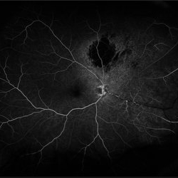
Ultra-wide field fluorescein angiography of a 57-year-old male s/p I-125 brachytherapy for malignant melanoma affecting his right eye. The patient’s radiation retinopathy has resulted in retinal vascular occlusive disease and optic nerve edema.
Photographer: Olivia Rainey
Imaging device: Optos
Condition/keywords: branch retinal vein occlusion (BRVO), fluorescein angiogram (FA), I-125 brachytherapy, malignant melanoma, optic disc edema, Optos, radiation retinopathy, ultra-wide field imaging
-
 Radiation Retinopathy; BRVO with Macular Edema
Apr 26 2023 by Denica Rodriguez
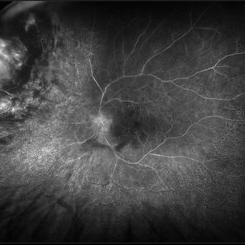
Ultra-wide field fluorescein angiography of a 61 year old male with radiation retinopathy following brachytherapy for choroidal melanoma of his left eye. Following treatment, patient developed a branch retinal vein occlusion both ischemic and non-ischemic. Anti-VEGF injections were recommended. The fine needle biopsy showed a class 2 uveal melanoma. Patient also has diabetic retinopathy affecting both eyes. Patient's vision at the time the image was taken was Dcc 20/80-1.
Photographer: Denica Rodriguez COA, ST
Imaging device: Optos California
Condition/keywords: branch retinal vein occlusion (BRVO), Choroidal melanoma, diabetic retinopathy, FA, fluorescein angiogram (FA), I-125 brachytherapy, macular edema, melanoma, Optos, radiation retinopathy, Retina, ultra-wide field imaging
-
 Regressed Iridociliary Medulloepithelioma (Status Post Plaque Brachytherapy)
Feb 12 2020 by Scott D Walter, MD, MSc, FASRS
2-year-old male with regressed iridociliary medulloepithelioma, one month status post plaque brachytherapy.
Condition/keywords: I-125 brachytherapy
-
 Choroidal Melanoma
Mar 26 2024 by Xitlali Caterina
Ultra-widefield fundus photograph of a 40-year-old woman with Choroidal Melanoma in right eye. Patient present with 20/50+2 vision in the right eye. Patient reported having frequent headaches located frontal area of their head and sometimes radiated to the right side as well. Patient also noted eye pain in both eyes that has remained constant for many years, as well as light sensitivity. The physician stated that since this is a medium-sized tumor, the treatment options include I-125 brachytherapy or enucleation. He recommended I-125 brachytherapy.
Photographer: Xitlali Caterina
Imaging device: Optos California RGB
Condition/keywords: fundus photography, Optos, OPTOS CALIFORNIA, superior retina, ultra-wide field imaging, ultra-widefield image
 Loading…