Search results (436 results)
-
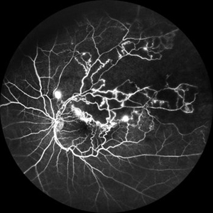
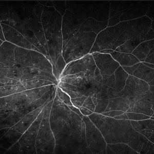
 Active Proliferative Diabetic Retinopathy
Jul 12 2024 by Korey Starkey
Fluorescein angiogram performed on 35 year old female with active proliferative diabetic retinopathy. Patient has peripapillary vascular loop and history of PRP treatment in both eyes. Patients left eye vision measured at Dcc20/200-1 at this visit.
Photographer: Korey Starkey
Imaging device: Optos
Condition/keywords: FLUORESCEIN ANGIOGRAPHY, hyperfluorescence, laser scarring, Optos, proliferative diabetic retinopathy (PDR), sea fan, ultra-wide field imaging, vascular loop
-
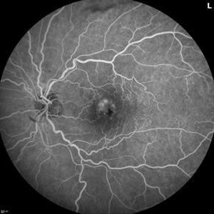
 Branch Retinal Vein Occlusion
Jul 23 2025 by Malvika Singh
Fluorescein angiogram of a 52 year old man showing capillary non perfusion areas and leakages along the superotemporal arcade and at the macula.
Photographer: Dr Malvika Singh, Retina Foundation, Ahmedabad, India
Imaging device: Mirante SLO/OCT
Condition/keywords: branch retinal vein occlusion (BRVO), CNP areas, FLUORESCEIN ANGIOGRAPHY, fluorescein leakage, macular edema
-
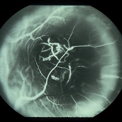
 Branch Retinal Vein Occlusion
Oct 17 2012 by Sharon Fekrat, MD FACS FASRS
Fluorescein angiography of an inferior perfused branch retinal vein occlusion in the left eye of a middle aged male with hypertension. The foveal avascular zone is irregular. Subretinal hemorrhage is present.
Photographer: John Reaves, Ophthalmic Photographer, Durham VA Medical Center Eye Clinic Imaging Suite, Durham, NC
Imaging device: Fluorescein Angiography
Condition/keywords: branch retinal vein occlusion (BRVO), subretinal hemorrhage
-
 Branch Retinal Vein Occlusion with Multifactorial Macular Edema and Epiretinal Membrane
Oct 3 2024 by Logan ryzenga
Fluorescein angiogram of a 62 year old woman with cystoid macular edema from concurrent Epiretinal Membrane and Branch Retinal Vein occlusion. She has an extensive history of anti-VEGF injections with stable but unresolved macular edema. Following angiography, it was determined that an epiretinal membrane peel would be indicated in an attempt to achieve resolution of macular edema.
Photographer: Logan Ryzenga
Imaging device: Heidelberg Spectralis
Condition/keywords: 55-degrees, branch retinal vein occlusion (BRVO), cystoid macular edema (CME), epiretinal membrane (ERM), Fluorescein angiography, heidelberg spectralis, hyperfluorescence, leakage, left eye, OS, wide angle imaging
-
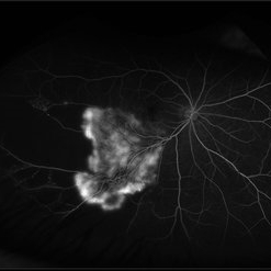 Branch Retinal Vein Occlusion with Retinal Neovascularization
Mar 21 2024 by Isaac Agranoff
Fundus angiography photograph of a 63 year old male presenting with worsening blurry vision OD for 4 years with new transient floaters (vision 20/160 PH 20/60). Fluorescein angiography revealed significant capillary non-perfusion corresponding to the area, with peripheral vascular remodeling. Physician recommended anti-VEGF therapy and FA-guided supplemental PRP given the size of the NVE.
Photographer: Isaac Agranoff
Imaging device: Optos California
Condition/keywords: branch retinal vein occlusion (BRVO), EYLEA, FLUORESCEIN ANGIOGRAPHY, Neovascularisation elsewhere (NVE), Optos
-
 Central Retinal Artery Occlusion Fluorescein Angiography
Sep 25 2024 by Gustavo Uriel Fonseca Aguirre
43 year-old female with history of rheumatoid arthritis, fluorescein angiography in central retinal artery occlusion.
Photographer: Gustavo U. Fonseca Aguirre, Fundación Hospital Nuestra Señora de la Luz, Ciudad de México
Condition/keywords: central retinal artery occlusion, Fluorescein angiography
-
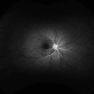
 Central Retinal Vein Occlusion
Sep 27 2024 by Korey Starkey
Fluorescein angiogram of a 75 year old patient with central retinal vein occlusion. FA shows areas of patchy ischemia and petaloid leakage. Patient is being treated with anti-vegf treatments at this time.
Photographer: Korey Starkey
Condition/keywords: central retinal vein occlusion (CRVO), FLUORESCEIN ANGIOGRAPHY, ischemia, macular edema, petaloid leakage, ultra-widefield image
-
 Central Retinal Vein Occlusion With Waldenstroms macroglobulinemia
Jun 18 2025 by Korey Starkey
64-year-old patient presents with CRVO with secondary macular edema in both eyes. Venous beading present in 2/4 quadrants OU. Patient diagnosed with Waldenstroms macroglobulinemia, found on SPEP and bone marrow biopsy. Treatment recommended of anti-vegF intravitreal injections OU.
Photographer: Korey Starkey
Imaging device: Optos
Condition/keywords: attenuated vessels, central retinal vein occlusion (CRVO), CRVO, FA early phase, FLUORESCEIN ANGIOGRAPHY, macular edema, Optomap, OPTOS CALIFORNIA, severe NPDR, venous beading, Waldenstroms macroglobulinemia
-
 Choroidal Hemangioma
Jul 30 2024 by Korey Starkey
ICG image of a77 year-old female with choroidal hemangioma. The physician states the hypercyanesence in the right eye is consistent with hemangioma but no typical late washout observed. He also notes high internal reflectivity make hemangioma possible. Patients vision at time of imaging VA OD: sc20/200 PH20/60-1; plan to follow patient at 6 month intervals at this time.
Photographer: Korey Starkey
Imaging device: Optos
Condition/keywords: Choroidal Hemangioma, Fluorescein angiography, indocyanine green (ICG) angiography, Optos
-
 Choroidal Hemangioma 4 Ways
Mar 13 2025 by Virginia Gebhart
Color fundus, FAF, late FA, late ICG of 64 year old male with choroidal hemangioma. Early hyperfluorescence with late leakage on FA, early hypercyanescence with late washout (25 min) on ICG.
Photographer: Virginia Gebhart, Retina Consultants of Carolina
Imaging device: Optos California
Condition/keywords: autofluorescence imaging, choroidal hemangioma, FA late phase, Fluorescein angiography, hemangioma, indocyanine green (ICG) angiography
-
 Choroidal Melanoma
Oct 27 2023 by Virginia Gebhart
76 year old male with suspicious pigmented choroidal lesion with new collar button growth. Blocking defect and vascularity noted on FA
Photographer: Virginia Gebhart
Condition/keywords: FA late phase, fluorescein angiogram (FA), Fluorescein angiography, melanoma
-
 Choroidal Melanoma
Jan 4 2024 by Virginia Gebhart
57 year old female with new choroidal melanoma. Early hyperfluorescence with vascularity and minimal late leakage on FA.
Photographer: Virginia Gebhart
Imaging device: Optos California
Condition/keywords: FA, FA early phase, fluorescein angiogram (FA), Fluorescein angiography
-
 Choroidal Melanoma
May 28 2014 by Henry J. Kaplan, MD
Fluorescein angiography of a patient with choroidal melanoma clearly shows the double circulation of the retina and whithin the melanoma #2
Imaging device: Fluorescein angiography
Condition/keywords: melanoma
-
 Choroidal Melanoma FA
Nov 14 2023 by Virginia Gebhart
Fluorescein angiogram of 69 year old male with small lesion consistent with choroidal melanoma. Small pigmented elevated choroidal lesion just below ON with drusen, RPE changes and trace questionable OP present in the left eye. Extensive imaging and ultrasound was performed for further evaluation and documentation.
Photographer: Virginia Gebhart
Imaging device: Optos
Condition/keywords: fluorescein angiogram (FA), Fluorescein angiography
-
 Chronic CRVO
Dec 12 2024 by Korey Starkey
Fluorescein Angiography of a 62 year-old man with chronic central retinal vein occlusion. Vision is 20/200.
Photographer: Korey Starkey
Imaging device: Optos
Condition/keywords: capillary nonperfusion, central retinal vein occlusion (CRVO), FLUORESCEIN ANGIOGRAPHY, ischemia, microaneurysms, Optos
-
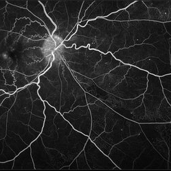 Diabetic Retinopathy
Nov 20 2024 by Korey Starkey
64 year old female being monitored for moderate-severe diabetic retinopathy.
Photographer: Korey Starkey
Condition/keywords: capillary nonperfusion, FA, FLUORESCEIN ANGIOGRAPHY, microaneurysms, nonproliferative diabetic retinopathy, Optos, OPTOS CALIFORNIA, tortuous vessels
-
 FA/ICG Choroidal Melanoma
Mar 10 2025 by Virginia Gebhart
Side by Side comparison of late FA/ICG on choroidal melanoma. FA showed early lacy hyperfluorescence with late leakage, ICG showed late Hypocyanescence.
Photographer: Virginia Gebhart, Retina Consultants of Carolina
Imaging device: Optos California
Condition/keywords: FA, Fluorescein angiography, fluorescein leakage, indocyanine green (ICG) angiography
-
 Fluorescein and Indocyanine Green Angiography in Right Eye in Case of Choroidal Hemangioma
Nov 29 2024 by Anand Temkar
Right eye Fluorescein and Indocyanine green angiography of a 42 year old male in case of Choroidal hemangioma. Choroidal hemangioma have a unique pattern of circulation where the large blood vessels produce a “COARSE VASCULAR PATTERN.” Fluorescein angiography of circumscribed choroidal hemangiomas typically reveals very early hyperfluorescence of larger-caliber choroidal blood vessels either before or simultaneously with the initial filling of the retinal arterioles. Indocyanine green angiography typically shows filling of the intralesional vascular channels, intense hypercyanescence of the lesion by the intermediate frames (peaks around 3-4 minutes) and late washout of the central portion of the lesion.
Photographer: Dr.Anand Temkar- Retina Foundation, Ahmedabad
Imaging device: Mirante
Condition/keywords: Choroidal Hemangioma, FLUORESCEIN ANGIOGRAPHY, indocyanine green (ICG) angiography
-
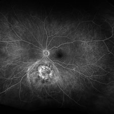
 Focal Chorioretinitis
Jul 11 2024 by Virginia Gebhart
67 year old female with punched-out CR scars. Hx of laser 3x for apparent peripapillary CNV. ESR, CRP, toxo, IgG/IgM all "normal." Bartonella, quant gold, and FTA-ABS ordered given possibility of neuroretinitis. Vision CF
Photographer: Virginia Gebhart
Imaging device: Optos California
Condition/keywords: FA, fluorescein angiogram (FA), FLUORESCEIN ANGIOGRAPHY, focal chorioretinitis, optic neuritis
-
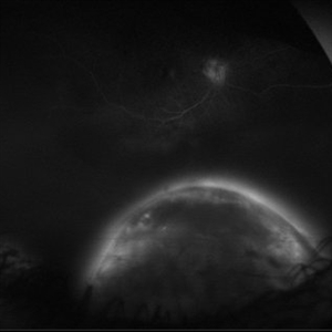 Full Moon
Aug 20 2025 by Gustavo Uriel Fonseca Aguirre
This ultra-widefield fluorescein angiography reveals a hyperfluorescent peripheral inferior choroidal melanoma. The lesion demonstrates early heterogeneous hyperfluorescence with progressive late staining and diffuse leakage.
Photographer: Gustavo U. Fonseca Aguirre, Hospital Conde de Valenciana, Ciudad de México
Condition/keywords: choroidal melanoma, FLUORESCEIN ANGIOGRAPHY
-
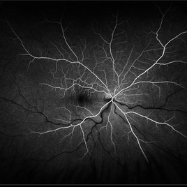 Hemiretinal Vein Occlusion
Nov 14 2024 by Brandon I Fram, MD
40 year-old male with vision changes and observed hemiretinal vein occlusion.
Condition/keywords: branch retinal vein occlusion (BRVO), fluorescein angiogram (FA), Fluorescein angiography, hemi CRVO, hemicentral retinal vein occlusion
-
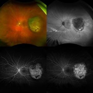 Melanoma Multimodal Evaluation
Feb 10 2025 by Gustavo M. Hüning, MD, MBA, FASRS
UWF multimodal imaging of an 37-year-old woman with a choroidal melanoma. The mosaic shows a colored retinography; a FAF with regions of previous serous detachments; an early stage of angiography and a later time.
Photographer: Gustavo M. Hüning, HÜNING Clínica do Olhar, Santa Maria - Brazil
Imaging device: Optos California
Condition/keywords: Autofluorescence, Choroidal, Fluorescein angiography, melanoma, multimodal imaging, ultra-wide field imaging
-
 Neovascularization in Posterior Uveitis
Jul 27 2023 by Zach Seim
An ultra-widefield fluorescein angiogram of a 72 year old male with Posterior Uveitis and Neovascularization affecting the right eye. Patient's vision at the time of the image was Dcc 20/25. Dr. Korot states that the fluorescein angiogram shows patchy leakage throughout both eyes, with peripheral nonperfusion and secondary neovascularization. The patient was asked to get an extensive serological workup in an effort to identify any systemic autoimmune or infectious etiology as the cause for their severe inflammation.
Photographer: Zach Seim
Imaging device: OPTOS California
Condition/keywords: fluorescein angiogram (FA), FLUORESCEIN ANGIOGRAPHY, fluorescein leakage, neovascularization (NV), Optos, OPTOS CALIFORNIA, posterior uveitis, right eye, ultra-wide field imaging, ultra-widefield image
-
 Occlusive Retinal Vasculitis
Oct 3 2024 by Logan ryzenga
4 view ultra-widefield Optos fluorescein angiogram in the left eye of a 39 year old woman occlusive retinal vasculitis with four quadrant Kyrieleis plaques. This is a showcase of a suspected, rarely reported, and atypical presentation of Behcet's Syndrome.
Photographer: Logan Ryzenga
Imaging device: Optos California
Condition/keywords: Behcet's Disease, Behcet's uveitis, Fluorescein angiography, fluorescein leakage, kyrieleis plaques, non-perfusion, OPTOS, OPTOS CALIFORNIA, ultra-wide field imaging, Uveitis
-
 Ocular Ischemic Syndrome
Jun 27 2023 by Mauricio Bayram-Suverza, MD
Fundus photograph of a 72-year-old man referred to the retina department due to long-term, painless vision loss in his left eye. Following fluorescein angiography, prolonged arteriovenous transit time and hypoperfusion were detected. Consequently, the patient was advised to undergo cardiological evaluation.
Photographer: Mauricio Bayram-Suverza, Fundación Hospital Nuestra Señora de la Luz
Imaging device: Optos California
Condition/keywords: Fluorescein angiography, ocular ischemic syndrome, Ultra-wide field retinal imaging
 Loading…