Search results (52 results)
-
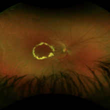
 Advanced coats disease
Dec 27 2023 by NIDHI PANWAR, MD FRCS Glasgow FNB FICO
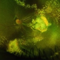
Fundus photograph of 6 year old otherwise healthy boy presented with right eye esotropia and poor vision with fundus picture depicting advanced exudative retinal disease suggestive of coats disease
Photographer: Nidhi Panwar, NMC Royal hospital, Sharjah , UAE
Condition/keywords: Coats disease, subretinal exudates
-
 All That Glows Yellow Isn’t Mellow: Coats' Disease Unveiled
Nov 4 2025 by SHRADDHA RAJ SHRIVASTAVA
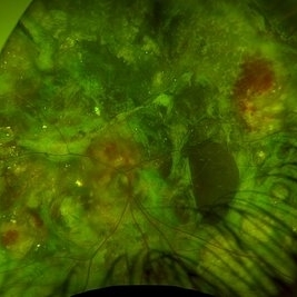
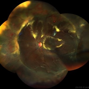
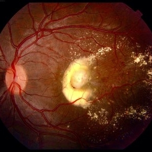
Montage fundus image of an 11 year old boy diagnosed with left eye Coats' disease (stage 3A1), reveals a hyperemic disc and surrounding intra-retinal exudates superior to the disc. There is a single fibroglial nodule at the macula causing submacular fibrosis with exudation. We can see areas of pigmentary changes and RPE atrophy in posterior pole and mid-peripheral retina supero-temporally. There is massive yellowish subretinal exudation in all the quadrants, which are associated with telangiectatic aneurysmal capillary dilation, more prominently seen in the nasal periphery. Supero-nasally we can also see an orange-red elevated vaso-proliferative mass with overlying dilated capillaries, which has likely developed secondary to untreated long standing disease. We can also see associated extrafoveal subtotal exudative retinal detachment in the inferior and nasal quadrants.
Photographer: Dr. Shraddha Raj Shrivastava
Imaging device: Nidek Mirante SLO/OCT (Confocal scanning/Spectral domain OCT)
Condition/keywords: COATS DISEASE, exudative detachment, leukocoria, subretinal exudates, Xanthocoria, yellow exudate
-
 Coats Disease
Sep 24 2024 by Gustavo Uriel Fonseca Aguirre
A 5-year-old male patient with no ophthalmological history, diagnosed with Coats disease in the right eye.
Photographer: Gustavo U. Fonseca Aguirre, Fundación Hospital Nuestra Señora de la Luz, Ciudad de México
Condition/keywords: Coats' disease
-
 Coats Disease
May 27 2025 by César Adrián Gómez Valdivia, MD
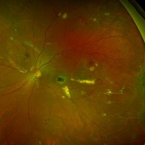
Fundus photograph of an 8 year-old male patient with Coats disease. Vascular leakage causes hard exudates which may be peripheral (near the vascular abnormalities) or midperipheral and central (at the macula). Findings were bilateral.
Photographer: @eyemissu2
Imaging device: California ICG OPTOS
Condition/keywords: Coats disease
-
 Coats Disease
May 27 2025 by César Adrián Gómez Valdivia, MD
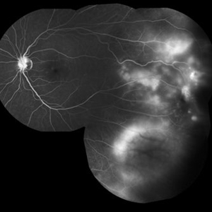
Fluorescein Angiography on an 8 year-old male patient with Coats disease. Vascular leakage causes hard exudates which may be peripheral (near the vascular abnormalities) or midperipheral and central (at the macula. Findings were bilateral.
Photographer: @eyemissu2
Imaging device: California ICG OPTOS
Condition/keywords: Coats disease
-
 Coats Disease
Sep 29 2024 by Tejaswita Verma
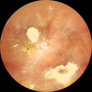
Fundus photo of the RE of a 14 y/o female ,nil premorbid presented with reduced vision in the RE ,diagnosed incidentally on ophthalmological examination elsewhere .Vision was finger counting 3 meters in the RE . Fundus picture reveals macular scar , subretinal and intraretinal exudation ,with scattered hemorrhages esp. in STQ, sclerosed vessels in superior, superonasal quadrant ,nasal, inferonasal quadrant, CR scars inferiorly, Telengiectatic vessels S/O Coat's disease. She was advised RE anti VEGF x1 + laser PRP + PST kenacort under GA with guarded prognosis.
Photographer: DR. TEJASWITA VERMA
Imaging device: MIRANTE
Condition/keywords: Coats' disease
-
 Coats disease
Jan 22 2023 by Mateus Queiroz Corrêa, MD
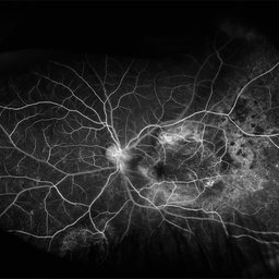
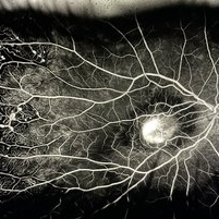
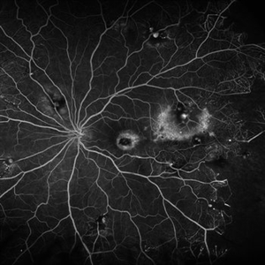
Ultra-widefield fluorescein angiography of a 11-year-old boy presenting temporal dilated and telangiectatic vessels associated non perfused areas. Macular leakage was also present showing vascular malformation in this region.
Photographer: Mateus Queiroz Correa, Sorocaba Eye Bank Hospital
Imaging device: California Optos, Ultra-widefield fluorescein angiography
Condition/keywords: COATS, Coats' disease
-
 Coats Disease
Mar 30 2024 by Karen Flores Guevara
Fundus photograph of a 9-year-old child with coats´ disease history of Scleral Buckling due to Retinal Detachment.
Photographer: Diana Elizabeth-Arellano-Acosta-MD Pediatric Retina,Asociación para Evitar la Ceguera en México IAP. México
Condition/keywords: Coats' disease
-
---thumb.jpg/image-square;max$300,300.ImageHandler) Coats Disease
Coats Disease
Oct 30 2012 by Lihteh Wu, MD
Fundus photograph of a 29-year-old man with no significant medical or ocular history. Patient complained of progressive loss of vision over the past few months. Notice the lipid exudation over the macula and the hyperplastic RPE.
Condition/keywords: hyperplastic retinal pigment epithelium (RPE), lipid exudation, retinal pigment epithelium
-
---thumb.jpg/image-square;max$300,300.ImageHandler) Coats Disease
Coats Disease
Oct 30 2012 by Lihteh Wu, MD
FA frame showing blocked fluorescence from the massive lipid exudation. There is also hyperfluorescence secondary to vascular leakage and hypofluorescence from the hyperplastic RPE. Superotemporal to the fovea there are areas of telangiectasia.
Condition/keywords: massive lipid exudation, retinal pigment epithelium, retinal telangiectasia
-
---thumb.jpg/image-square;max$300,300.ImageHandler) Coats Disease
Coats Disease
Oct 30 2012 by Lihteh Wu, MD
FA frame showing peripheral telangiectasia and some vascular leakage.
Condition/keywords: peripheral telangiectasia
-
 Coats Disease
Feb 7 2013 by Raj K. Maturi, MD
8-year-old male, flourescein angiogram, late phrase 4:22 minutes, OD.
Photographer: Stephan Morrow, Midwest Eye Institute Indianapolis Indiana
Imaging device: Heidelberg Spectralis
Condition/keywords: Spectralis
-
 Coats Disease
Feb 7 2013 by Raj K. Maturi, MD
8-year-old male, Heidelberg Spectralis OCT, OD.
Photographer: Stephan Morrow, Midwest Eye Institute Indianapolis Indiana
Imaging device: Heidelberg Spectralis
Condition/keywords: optical coherence tomography (OCT), Spectralis
-
 Coats Disease
Feb 7 2013 by Raj K. Maturi, MD
8-year-old male, flourescein angiogram, venous phrase, OD.
Photographer: Stephan Morrow, Midwest Eye Institute Indianapolis Indiana
Imaging device: Heidelberg Spectralis
Condition/keywords: Spectralis
-
 Coats Disease
Oct 9 2012 by Alan D. Letson, MD
Coats Disease
Photographer: Beverly Radcliffe
Condition/keywords: retinal macroaneurysm, retinal telangiectasia
-
---thumb.jpg/image-square;max$300,300.ImageHandler) Coats disease
Coats disease
Jan 11 2013 by Hyung-Woo Kwak, MD
Fundus imaging shows hemorrhage and hard exudates from leaking blood vessel.
Photographer: Taegi Kim, Kyung Hee Univsersity Hospital, Seoul
Imaging device: Zeiss f 450 plus
Condition/keywords: argon photocoagulation
-
 Coats Disease
Feb 18 2022 by Ahmad B. Tarabishy, MD
43 year old gentleman with poor vision in his left eye for many years. Examination shows multiple retinal telangiectasias and aneurysms. Ultrawide field fluorescein angiography shows light-bulb aneurysms, telangiectasias, and extensive vascular remodeling and non-perfusion.
Photographer: Dr. Angela Rico, Retina Specialists of Tampa
Condition/keywords: Coats' disease
-
 Coats Disease
Apr 16 2015 by Rita Couceiro, MD, MS
Fundus photograph and fluorescein angiography pictures of a 13-year-old girl with Coats Disease, showing abnormal telangiectatic vessels and intense exsudation in the inferior retinal periphery of the left eye.
Condition/keywords: Coats' disease, retinal telangiectasia
-
 Coats Disease
Jul 7 2022 by Gabriel Costa Andrade, PhD
Fundus photograph of a 31-year-old man with no medical or ocular history. Patient complained of progressive loss of vision over the past few months OS. Notice the lipid exudation over the macula and telangiectatic vessels.
Photographer: Dr Gabriel Andrade
Condition/keywords: Coats' disease
-
 Coats Disease
May 27 2016 by Olivia Rainey
Composite fluorescein angiogram of the left eye of a man with Coats Disease.
Photographer: Olivia Rainey
Imaging device: Heidelberg Spectralis
Condition/keywords: Coats' disease, composite, fluorescein angiogram (FA), fluorescein leakage, Heidelburg Spectralis
-
 Coats Disease
May 2 2023 by JEFFERSON R SOUSA, Tecg.º (Biomedical Systems Technology)
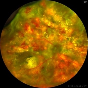
Male patient, 7 years old, with low acuity. The patient's difficulty seeing was noticed in a school eye care project. In the screening exam, the child's difficulty seeing was evident. In the specialty exams, important retinal alterations suggestive of Coats Disease were noted.
Photographer: JEFFERSON ROCHA DE SOUSA - Department of Retina at Institute Suel Abujamra - ISA, São Paulo - Brazil.
Imaging device: Clarus 700 - Zeiss.
Condition/keywords: Coats disease
-
 Coats Disease
May 23 2024 by ARVIND JAIN M
a.right eye fundus image and b. FFA montage of a 8 year old boy showing light bulb aneurysms of the arterioles with exudation with sub retinal fibrosis and telangiectasia in periphery who complained of defective vision, classical of coats disease.
Photographer: Dr. Arvind Jain M, MBBS,MS Ophthal, FVRS
Condition/keywords: COATS DISEASE, Leber's miliary aneurysm, light-bulb aneurysms
-
 Coats Disease
Aug 25 2022 by Maxwell J Wingelaar, MD
A 12-year-old male with Coats' disease
Photographer: Jarrod Wehmeier
Condition/keywords: Coats' disease
-
 Coats disease
Nov 8 2022 by Heitor Nogueira
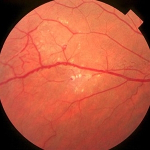
Fundus photograph of an 12-year-old asymptomatic patient. It is possible to observe the presence of vascular telangiectasias associated with areas of exudation without the presence of a tumor lesion.
Photographer: Heitor Nogueira, Instituto Penido Burnier, Campinas-SP, Brazil
Condition/keywords: Coats' disease
 Loading…