Search results (7 results)
-
 Anemic Retinopathy in a Young Female
Jan 15 2022 by KRISHNENDU NANDI, MS
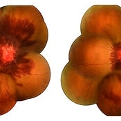
Fundus photograph of a 29-year-old female presented with retinal hemorrhages in both eyes with decrease in vision for 1 month. Hemoglobin level was 5.9gm/dl, suggestive of anemic retinopathy in both eyes.
Photographer: Krishnendu Nandi, Netralayam Eye Care Centre, Kolkata, India
Imaging device: Topcon
Condition/keywords: anaemic retinopathy, anemic retinopathy, retinal hemorrhage
-
 Roth Spots Everywhere
Apr 23 2025 by Thirumalesh Mochi Basavaraj, MD
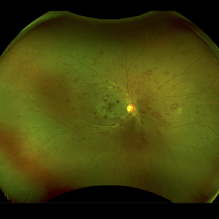
Fundus image of a 39 year-old female with symptoms of blurring of vision , who was severely anemic who was myelodysplastic on bone marrow aspiration cytology.
Photographer: Vivekananda
Imaging device: Optos Daytona
Condition/keywords: ANEMIC RETINOPATHY, MYELODYSPLATIC RETINOPATHY, Roth spots
-
 Thrombocytopenia
Sep 24 2024 by DR Rohit Gupta
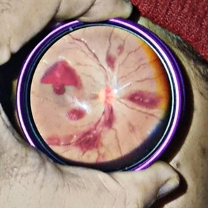
Fundus photography of a 16 year old female suffering from severe thrombocytopenia. On fundus examination, multiple roth spots and subhyaloid hemorrhage were seen.
Photographer: Dr Rohit gupta
Imaging device: Samsung S21
Condition/keywords: ANEMIC RETINOPATHY, hemorrhage, leukemia, retinal hemorrhage, Roth spots, thrombocytopenia
-
 WAXING MOON OR WANING MOON?
Oct 12 2023 by Deepti A Kulkarni, M.B.B.S., D.N.B., F.V.R.
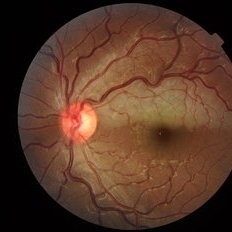
FUNDUS PHOTO OF AN 18 YEAR OLD. VISION AT PRESENTATION 6/6 WITH DIFFICULTY IN READING AND A BLACK SPOT WHEN TRYING TO FOCUS. ANAEMIC FOR OVER TWO YEARS - IN HER RECOVERY PHASE WHERE HEMOGLOBIN HAD RISEN FROM <5g/dL TO 7.5 g/dL WHERE SHE HAD REACTIVE THROMBOCYTOSIS CAUSING HYPERCOAGULABILITY AND RETINAL ARTERIOLAR OCCLUSION.
Photographer: DEEPTI KULKARNI, KULKARNI EYE HOSPITAL, MIRAJ, INDIA
Imaging device: TOPCON
Condition/keywords: ANEMIC RETINOPATHY, MACULAR BRANCH ARTERIAL OCCLUSION
-
 Anemic Retinopathy Related Retinal Hemorrhages
Nov 5 2019 by Chinmayi Vyas
Anemic retinopathy related retinal hemorrhages in a 24 years old male with Hb of 4.2gm/ dl. The manifestations of anemic retinopathy are nonspecific and may closely simulate hypertensive or diabetic retina. Retinal changes in anemia are cotton wool spots, venous tortuosity, and hemorrhages which may be present at all levels of the retina and choroid. All retinal hemorrhages can occur when Hb falls below 8 g/100 ml or if the platelet count falls below 50,000/cumm. The combination of severe anemia and thrombocytopenia is likely to produce retinal hemorrhages. The Roth’s spots or white centre hemorrhages are typically associated with bacterial endocarditis , anemia and other systemic conditions. The white center is suspected to represents focal ischemia, inflammatory or infectious infiltrate, fibrin or accumulation of neoplasticism cells.
Photographer: Dr Chinmayi Vyas
Condition/keywords: retinal hemorrhage
-
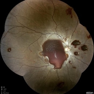 Anemic Retinopathy Related Retinal Hemorrhages
Nov 5 2019 by Chinmayi Vyas
Anemic retinopathy related retinal hemorrhages in a 24 years old male with Hb of 4.2gm/ dl. The manifestations of anemic retinopathy are nonspecific and may closely simulate hypertensive or diabetic retina. Retinal changes in anemia are cotton wool spots, venous tortuosity, and hemorrhages which may be present at all levels of the retina and choroid. All retinal hemorrhages can occur when Hb falls below 8 g/100 ml or if the platelet count falls below 50,000/cumm. The combination of severe anemia and thrombocytopenia is likely to produce retinal hemorrhages. The Roth’s spots or white centre hemorrhages are typically associated with bacterial endocarditis , anemia and other systemic conditions. The white center is suspected to represents focal ischemia, inflammatory or infectious infiltrate, fibrin or accumulation of neoplasticism cells.
Photographer: Dr Chinmayi Vyas, Nethradhama superspeciality eye hospital , banglore, india
Imaging device: Eidon fundus imaging
Condition/keywords: anaemic retinopathy
-
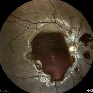
 Macular Hemorrhage Secondary to Anemic Retinopathy
Apr 18 2022 by Deepak Bhojwani, MS
Fundus image of a young 28 year old patient who has been diagnosed as 'PRIMARY BONE MARROW APLASIA' by hematologist showing large macular hemorrhage (sub -ILM Heme mound). Few Roth spots were also seen in midperiphery suggesting 'ANEMIC RETINOPATHY'.
Photographer: DEEPAK BHOJWANI
Condition/keywords: anaemic retinopathy, BONE MARROW APLASIA
 Loading…