Search results (17 results)
-
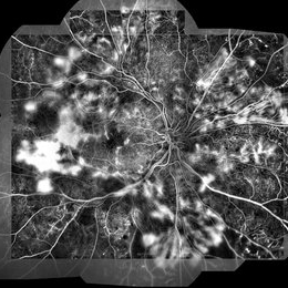
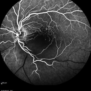 Central Retinal Artery Occlusion
May 16 2017 by Olivia Rainey
Fluorescein angiogram of an 66-year-old female with a central retinal artery occlusion affecting her left eye.
Photographer: Olivia Rainey
Imaging device: Heidelberg Spectralis
Condition/keywords: 50 degreescentral retinal artery occlusion (CRAO)fluorescein angiogram (FA)left eyemid phaseretinal ischemia
-
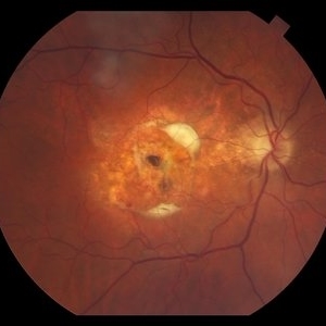 Disciform Scar Due to Exudative Macular Degeneration
Feb 2 2018 by Olivia Rainey
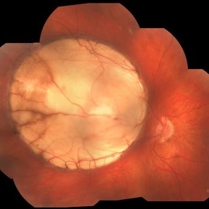
Color fundus photograph of a 74-year-old male with a central disciform scar due to exudative macular degeneration.
Photographer: Olivia Rainey
Imaging device: Topcon 50dx
Condition/keywords: 50 degreesage-related macular degeneration (AMD)central disciform scarcolor fundus photograph
-
 Hemangioma of Retina
Sep 11 2018 by Carolyn Daley
50 degree OCT imaging of a 20-year-old with multiple bilateral hemangiomas. Patient was diagnosed with Von Hippel-Lindau Syndrome.
Photographer: Carolyn Daley, Retina Specialists of Michigan
Imaging device: Heidelberg Spectralis
Condition/keywords: 50 degreesedemahemangiomaoptical coherence tomography (OCT)Von Hippel-Lindau
-
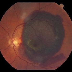 Massive Subretinal Hemorrhage Due to Exudative Macular Degeneration
Feb 2 2018 by Olivia Rainey
Color fundus photograph of a 74-year-old male with a massive subretinal hemorrhage due to exudative macular degeneration.
Photographer: Olivia Rainey
Imaging device: Topcon 50dx
Condition/keywords: 50 degreesage-related macular degeneration (AMD)color fundus photographleft eyesubretinalhemorrhage
-
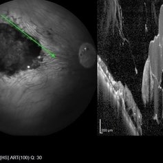 Retinal Cavernous Hemangioma
Oct 22 2020 by Olivia Rainey
Widefield OCT of a 31-year-old male presenting with a retinal cavernous hemangioma affecting his left eye. Patient was 18-years-old when he was diagnosed with a retinal cavernous hemangioma. He has had a few episodes of vitreous hemorrhages since then. His vision was 20/20-1 in both eyes.
Photographer: Becca Harris
Imaging device: Heidelberg Spectralis
Condition/keywords: 50 degreescavernous hemangioma of the retinaHeidelburg Spectralisleft eyeoptical coherence tomography (OCT)wide angle imaging
-
 Retinitis Pigmentosa
May 27 2016 by Olivia Rainey
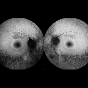
Bilateral fundus autofluorescence images of retinitis pigmentosa.
Photographer: Olivia Rainey
Imaging device: Heidelberg Spectralis
Condition/keywords: 50 degreesbilateralfundus autofluorescence (FAF)hereditary retinal dystrophyretinitis pigmentosa
-
 Arterial Occlusion With Preservation of Cilioretinal Network
Aug 29 2016 by JEFFERSON R SOUSA, Tecg.º (Biomedical Systems Technology)
Patient male, 52-years-old, denies hypertension, blood systemic, and diabetes. Refers low subtle of vision, field loss and stain central.
Photographer: JEFFERSON R SOUSA - Institute Dr. Suel Abujamra / São Paulo - Brazil
Imaging device: Acquisition of the image in the Camera background Topcon TRC-50 Dx - IA, Keystone field photo of 50 Degrees. Composition automatic of Imaginet with manual adjustment.
Condition/keywords: arterial occlusionhypertension
-
 BRVO and VMT Vitreo Macular Traction, Color Photo
Apr 18 2013 by James B. Soque, CRA, OCT-C, COA, FOPS
Color Photograph, 50 degrees, mag 2X of 79-year-old white female, VA sc 20/40, with BRVO OS, and VMT OS, diagnosed on exam and SD OCT. See accompanying RF and FA photos reveal BRVO IT OS, and SD OCT reveals VMT OS.
Photographer: James B. Soque, CRA, COA, Island Retina, Shirley, NY
Imaging device: Topcon TRC-50DX with MERGE software
Condition/keywords: branch retinal vein occlusion (BRVO)vitreomacular traction (VMT)
-
 BRVO and VMT Vitreo Macular Traction, FA Early Phase
Apr 18 2013 by James B. Soque, CRA, OCT-C, COA, FOPS
Early FA photo, 50 degrees, mag 2X of 79-year old white female, VA sc 20/40, with BRVO OS, and VMT OS, diagnosed on exam and SD OCT. See accompanying FC and RF photos reveal BRVO IT OS, and SD OCT reveal VMT OS.
Photographer: James B. Soque, CRA, COA, Island Retina. Shirley, NY
Imaging device: Topcon TRC-50DX with MERGE software
Condition/keywords: branch retinal vein occlusion (BRVO)vitreomacular traction (VMT)
-
 BRVO and VMT Vitreo Macular Traction, Red Free Photo
Apr 18 2013 by James B. Soque, CRA, OCT-C, COA, FOPS
Red Free Photo, 50 degrees, mag 2X of 79-year-old white female, VA sc 20/40, with BRVO OS, and VMT OS, diagnosed on exam and SD OCT. See accompanying FC and FA photos reveal BRVO IT OS, and SD OCT reveal VMT OS.
Photographer: James B. Soque, CRA, COA, Island Retina, Shirley, NY
Imaging device: Topcon TRC-50DX with MERGE software
Condition/keywords: branch retinal vein occlusion (BRVO)vitreomacular traction (VMT)
-
 Coloboma
Jan 23 2018 by JEFFERSON R SOUSA, Tecg.º (Biomedical Systems Technology)
Male patient, 22 years old, with low vision since infancy. In retinal and retinal mapping examinations, important alterations were observed in the formation of retinochoroidal structures suggestive of coloboma.
Photographer: JEFFERSON R SOUSA - Study Center and Ophthalmological Research Dr. Andre M V Gomes, Dr. Suel Abujamra Institute São Paulo-Brazil
Imaging device: Acquisition of the image in the Camera background Topcon TRC-50 Dx - IA, Keystone field photo of 50 Degrees. Composition automatic of Imaginet with manual adjustment
Condition/keywords: colobomacoloboma of choroid
-
 Diabetic Retinal Hemorrhages in Proliferative Diabetes
Sep 10 2012 by James B. Soque, CRA, OCT-C, COA, FOPS
Fundus Photo of Severe Proliferative Diabetic with Retinal Hemorrhages, Left eye, scattered laser treatment. View: 50 Degrees
Photographer: James Soque, CRA, COA, Island Retina, Shirley, NY
Imaging device: Topcon TRC 50 DX
Condition/keywords: proliferative diabetic retinopathy (PDR)
-
 Glaucoma
Feb 8 2018 by JEFFERSON R SOUSA, Tecg.º (Biomedical Systems Technology)
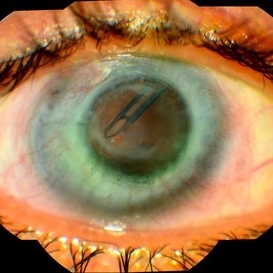
Male patient, 61-years-old in follow-up of glaucoma has several years. She performed trabeculectomy surgery with a tube implant.
Photographer: JEFFERSON R SOUSA - Study Center and Ophthalmological Research Dr. Andre M V Gomes, Dr. Suel Abujamra Institute São Paulo-Brazil
Imaging device: Fundus camera Acquisition of the image in the Camera background Topcon TRC-50 Dx - IA, field photo of 50 Degrees. Composition manual adjustment.
Condition/keywords: glaucoma
-
 Proliferative Diabetic Retinopathy
Jan 23 2018 by JEFFERSON R SOUSA, Tecg.º (Biomedical Systems Technology)
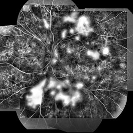
Male patient, 16-years-old, with type II diabetes mellitus, 320 MD / DL mean of glycemic control of the last five months. He attended the clinic complaining of low vision of both eyes. In the fluorescent retinal examination, several extravasation points corresponding to retinal neovascularization (Proliferative Diabetic Retinopathy) were observed.
Photographer: JEFFERSON R SOUSA - Study Center and Ophthalmological Research Dr. Andre M V Gomes, Dr. Suel Abujamra Institute São Paulo-Brazil
Imaging device: Acquisition of the image in the Camera background Topcon TRC-50 Dx - IA, Keystone field photo of 50 Degrees. Composition automatic of Imaginet with manual adjustment.
Condition/keywords: proliferative diabetic retinopathy (PDR)
-
 Proliferative Diabetic Retinopathy
Jan 23 2018 by JEFFERSON R SOUSA, Tecg.º (Biomedical Systems Technology)
Male patient, 16 years old, with type II diabetes mellitus, 320 MD / DL mean of glycemic control of the last five months. He attended the clinic complaining of low vision of both eyes. In the fluorescent retinal examination, several extravasation points corresponding to retinal neovascularization (proliferative diabetic retinopathy) were observed.
Photographer: JEFFERSON R SOUSA - Study Center and Ophthalmological Research Dr. Andre M V Gomes, Dr. Suel Abujamra Institute São Paulo-Brazil
Imaging device: Acquisition of the image in the Camera background Topcon TRC-50 Dx - IA, Keystone field photo of 50 Degrees. Composition automatic of Imaginet with manual adjustment.
Condition/keywords: proliferative diabetic retinopathy (PDR)
-
 Stereo View of Retinal Angiomatous Proliferation in Age-Related Macular Degeneration
Jan 21 2016 by James B. Soque, CRA, OCT-C, COA, FOPS
Stereo pair of 75-year-old white male with classic SRN with RAP lesion of right eye, actively receiving anti-VEGF treatment. 50 Degrees, no mag, L and R stereo pair. Single View of OD also visible in this case.
Condition/keywords: age-related macular degeneration (AMD)anti-VEGFretinal angiomatous proliferation (RAP)stereo pairsubretinal neovascularization (SRNV)
-
 Traumatic perforation and PVR
May 19 2022 by ALLAN GOMES DA SILVA
PVR, retinal detachment and macular dragging after iatrogenic traumatic perforation during peribulbar block. We can observe the inferior perforation, the brutal formation of PVR and the fovea being pulled.
Photographer: Edimilson Ferreira da Silva
Imaging device: Topcon TRC-50 DX, Imaginet 4.0, angle - 50 degrees
Condition/keywords: posterior perforationproliferative vitreoretinopathy (PVR)
 Loading…