Search results (14 results)
-
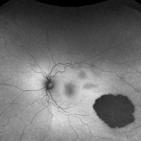
 White Without Pressure
Aug 23 2012 by Gerardo Garcia-Aguirre, MD
Photograph of the temporal peripheral retina showing an area of pale retina (white without pressure).
Photographer: Noemí Hernández, Asociación para Evitar la Ceguera en México
Imaging device: Zeiss FF4
Condition/keywords: pale retina, white without pressure
-
 White Without Pressure
Jan 31 2018 by Olivia Rainey
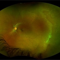
Ultra-wide field pseudocolor photograph of a 57-year-old female with white without pressure affecting her left eye. Patient will be having bloodwork done to rule out possible sarcoidosis or sickle cell.
Photographer: Olivia Rainey
Imaging device: Optos
Condition/keywords: blot hemorrhages, color fundus photograph, left eye, Optos, ultra-wide field imaging, white without pressure
-
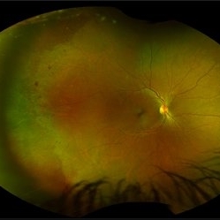
 White Without Pressure
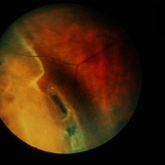
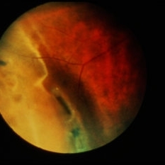
Mar 13 2020 by Anfisa Ayalon, MD
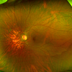
Fundus photograph of a 26-year-old woman with high myopia. Note inferotemporally margins of sharply demarcated WWP area.
Photographer: Anfisa Ayalon, MD., Meir Medical Center, Kfar Saba, Israel.
Imaging device: California, Optos 200 DTX
Condition/keywords: myopia, white without pressure
-
 Myopia with Lattice Degeneration and White Without Pressure in the Setting of Marfan's Syndrome
Aug 31 2020 by Sophia El Hamichi, MD
A 1-year-old female with Marfan's syndrome, myopia OU, congenital nystagmus and exotopia OD. Ultra-wide field imaging of her left eye showed lattice degeneration with atrophic retinal holes temporally, in addition to multiple sections of white without pressure.
Imaging device: Optos
Condition/keywords: atrophic retinal hole, lattice degeneration, Marfan's syndrome, myopia, Optos, ultra-wide field imaging
-
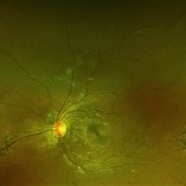
 Congenital Hypertrophy of the Retinal Pigment Epithelium Wide Field Optomap
Sep 24 2019 by Sophia El Hamichi, MD
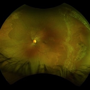
A 52-year-old female followed for 2 temporal lesions of CHRPE OD and white without pressure.
Photographer: Sophia El Hamichi,MD, Murray Ocular Oncology and Retina, Miami
Condition/keywords: congenital hypertrophy of the retinal pigment epithelium (CHRPE), Optomap, ultra-wide field imaging, white without pressure
-
 Ultra-Wide Field Fundus Photography Showing Lattice Degeneration
Mar 22 2021 by Sophia El Hamichi, MD
Lattice degeneration, atrophic holes, white without pressure OS in a 19-year-old female.
Condition/keywords: atrophic retinal hole, Optos, peripheral lattice degeneration, ultra-wide field imaging, white without pressure
-
 White Without Pressure/Dot Blot Hemorrhages
Jan 31 2018 by Olivia Rainey
Ultra-wide field pseudocolor photograph of a 57-year-old female with white without pressure affecting her right eye. Patient will be having bloodwork done to rule out possible sarcoidosis or sickle cell.
Photographer: Olivia Rainey
Imaging device: Optos
Condition/keywords: blot hemorrhages, Optos, ultra-wide field imaging
-
 Congenital Hypertrophy of the Retinal Pigment Epithelium Autofluorescence Optomap
Sep 24 2019 by Sophia El Hamichi, MD
A 52-year-old female followed for 2 temporal lesions of CHRPE OD and white without pressure.
Photographer: Sophia El Hamichi, MD, Murray Ocular Oncology and Retina, Miami
Condition/keywords: autofluorescence imaging, congenital hypertrophy of the retinal pigment epithelium (CHRPE), Optomap, ultra-wide field imaging, white without pressure
-
 Perivascular Bone Spicule Changes
Mar 1 2021 by Sophia El Hamichi, MD
A 19-year-old female African-American, who is followed for lattice degeneration and bone spicule changes OU. VA 20/20 OU. The bone spicule changes are stable throughout her follow-ups
Condition/keywords: bone spicule, lattice degeneration, Optos, perivascular, white without pressure
-
 White Without Pressure and Peripheral Retinoschisis
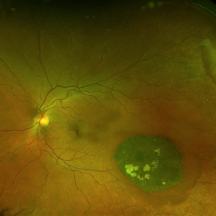
Dec 29 2022 by Gulnara Islamova
Fundus Photograph and OCT scan of an 18 year-old male with peripheral retinoschisis combined with WWOP lessions .Vitreoretinal traction is not visualized
Photographer: Gulnara Islamova, CENTER ZRENIYA Medical Clinic, LLC, Chelyabinsk, Russian Federation
Imaging device: Optovue XR Avanti
Condition/keywords: peripheral retinal degeneration
-
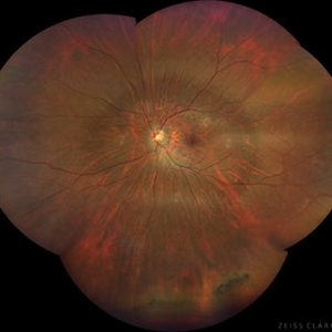
 Lattice Degeneration
Jan 5 2015 by H. Michael Lambert, MD
Lattice degeneration and white without pressure (stereo pair A).
Condition/keywords: lattice degeneration
-
 Pigmented Paravenous Chorioretinal Atrophy
Nov 5 2019 by Veronica A. Kon Graversen, MD
Fundus photograph of a 18-year-old African American with pigment and chorioretinal atrophy distributed along the retinal veins nasally and temporal. There is evidence of white without pressure.
Photographer: Alex Romera, Murray Ocular Oncology and Retina
Imaging device: Optos
Condition/keywords: pigmented paravenous chorioretinal atrophy (PPCRA)
-
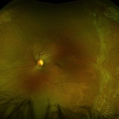
 High Myopia with Posterior staphyloma
Nov 7 2023 by Harsh Vardhan Singh, MS
27-year old with both eyes high myopia & posterior staphyloma with left eye peripheral lattice degeneration & white without pressure
Photographer: Harsh Vardhan Singh
Imaging device: Clarus 700
Condition/keywords: lattice degeneration, myopia, peripheral lattice degeneration, posterior staphylomaloma, white without pressure
-
 Lattice Degeneration
Jan 5 2015 by H. Michael Lambert, MD
Lattice degeneration and white without pressure (stereo pair B).
Condition/keywords: lattice degeneration
 Loading…