Search results (102 results)
-
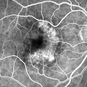
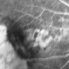
 Active Neovascular AMD With Disciform Scar
Apr 30 2015 by Mitzy E Torres Soriano, MD
Active neovascular AMD with disciform scar.
Photographer: Mitzy E. Torres Soriano, MD; Centro medico Cagua-Estado Aragua. Venezuela
Imaging device: TOPCON
Condition/keywords: disciform scar, disciform with hemorrhage, neovascular age-related macular degeneration (AMD), wet age-related macular degeneration (wet AMD)
-
 Advanced Wet AMD
Jan 29 2014 by Mallika Goyal, MD
OCT of the right eye of a 77-year-old patient with bilateral advanced AMD shows massive RPE detachment and sub sensory retinal fluid at presentation in January 2011.
Photographer: Mallika Goyal, MD, Apollo Health City, Hyderabad, India
Condition/keywords: wet age-related macular degeneration (wet AMD)
-
 Advanced Wet AMD
Jan 29 2014 by Mallika Goyal, MD
OCT of the right eye of a 77-year-old patient with bilateral advanced AMD shows regressing RPE detachment 1 year after initiating anti-VEGF therapy.
Photographer: Mallika Goyal, MD, Apollo Health City, Hyderabad, India
Condition/keywords: wet age-related macular degeneration (wet AMD)
-
---thumb.JPG/image-square;max$300,300.ImageHandler) Advanced wet AMD
Advanced wet AMD
Dec 1 2013 by Mallika Goyal, MD
Right eye of a 77 year old male patient with a large RPED, sub neurosensory retinal fluid and submacular bleed following 10 months gap in anti-VEGF therapy.
Photographer: Mallika Goyal, MD, Apollo Hospitals, Hyderabad, India
Condition/keywords: wet age-related macular degeneration (wet AMD)
-
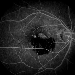
 Occult Wet AMD Fluorescein Angiogram
Jul 25 2014 by James B. Soque, CRA, OCT-C, COA, FOPS
FA OD image, early phase, 32 seconds, of 82-year-old white female with occult disease OD, anti-VEGF therapy since 2012, and retains 20/63 vision.
Photographer: James Soque, CRA COA
Imaging device: Topcon TRC 50 DX, OIS 5 MP Camera, MERGE software
Condition/keywords: wet age-related macular degeneration (wet AMD)
-
 Subretinal Hemorrhage Due to SRNVM, Fluorescein Angiogram Photograph
Dec 1 2016 by James B. Soque, CRA, OCT-C, COA, FOPS
89-year-old white male with NVAMD and new subretinal hemorrhage, fluorescein angiogram, early phase, of the right eye. Currently receiving anti VEGF treatment.
Photographer: James Soque, CRA, OCT-C, COA, Island Retina, Shirley, NY
Imaging device: Topcon TRC 50 DX, with MERGE software
Condition/keywords: hemorrhage, Hot spot, neovascular age-related macular degeneration (AMD), subretinal hemorrhage, subretinal blood, wet age-related macular degeneration (wet AMD)
-
 Wet AMD
Aug 12 2015 by Jared Watson
Wet AMD OD, receiving Avastin Q 1 month, since 2013, now with large subretinal hemorrhage.
Photographer: Jared Watson COT
Condition/keywords: wet age-related macular degeneration (wet AMD)
-
 Choroidal Hemorrhage, Subretinal Hemorrhage
Dec 18 2017 by Nichole Lewis
Choroidal hemorrhage, Subretinal Hemorrhage, wet macular degeneration,
Photographer: Nichole Lewis
Condition/keywords: choroidal hemorrhage, choroidal neovascularization (CNV), exudative age-related macular degeneration, subretinal hemorrhage, wet age-related macular degeneration (wet AMD)
-
---thumb.JPG/image-square;max$300,300.ImageHandler) Advanced Wet AMD
Advanced Wet AMD
Jan 29 2014 by Mallika Goyal, MD
Right eye of a 77-year-old male patient with bilateral advanced wet AMD shows regressing RPE detachment following 3 years of anti-VEGF therapy.
Photographer: Mallika Goyal, MD, Apollo Health City, Hyderabad, India
Condition/keywords: wet age-related macular degeneration (wet AMD)
-
 Advanced Wet AMD
Jan 29 2014 by Mallika Goyal, MD
OCT of the right eye of a 77-year-old patient with bilateral advanced AMD shows regressing RPE detachment 2 years after initiating anti-VEGF therapy.
Photographer: Mallika Goyal, MD, Apollo Health City, Hyderabad, India
Condition/keywords: wet age-related macular degeneration (wet AMD)
-
---thumb.JPG/image-square;max$300,300.ImageHandler) Advanced Wet AMD
Advanced Wet AMD
Jan 29 2014 by Mallika Goyal, MD
Right eye of a 77-year-old male patient with bilateral advanced wet AMD shows regressing RPE detachment following 3 years of anti-VEGF therapy.
Photographer: Mallika Goyal, MD, Apollo Health City, Hyderabad, India
Condition/keywords: wet age-related macular degeneration (wet AMD)
-
 Advanced Wet AMD
Jan 29 2014 by Mallika Goyal, MD
OCT of the right eye of a 77-year-old patient with bilateral advanced AMD shows regressing RPE detachment 3 years after initiating anti-VEGF therapy.
Photographer: Mallika Goyal, MD, Apollo Health City, Hyderabad, India
Condition/keywords: wet age-related macular degeneration (wet AMD)
-
 Subretinal Hemorrhage Due to SRNVM, Color Photograph
Dec 1 2016 by James B. Soque, CRA, OCT-C, COA, FOPS
89-year-old white male with NVAMD and new subretinal hemorrhage, 50 degree, color photograph of the right eye. Currently receiving anti VEGF treatment.
Photographer: James Soque, CRA- OCT-C, COA, Island Retina, Shirley, NY
Imaging device: Topcon TRC 50 DX, MERGE software
Condition/keywords: blood, choroidal neovascularization (CNV), color photo, subretinal blood, subretinal hemorrhage, wet age-related macular degeneration (wet AMD)
-
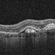 Sub-RPE Cholesterol Crystals in Wet AMD - Onion Sign
Sep 18 2017 by Theodore Leng, MD, MS, FASRS
Sub-RPE cholesterol crystals in Wet AMD - onion sign.
Imaging device: Cirrus
Condition/keywords: cholesterol crystals, crystals, onion sign, wet age-related macular degeneration (wet AMD)
-
 Occult ARMD, Color Fundus Photograph
May 17 2016 by James B. Soque, CRA, OCT-C, COA, FOPS
86-year-old white female after anti veg-f therapy. Color fundus photo of occult SRN with pigment mottling of the right eye.
Photographer: James B Soque, CRA, OCT-C, COA, Island Retina, Shirley, NY
Imaging device: Topcon TRC 50 EX, OIS 5 MP Camera, MERGE software
Condition/keywords: after treatment, color fundus photograph, occult, occult choroidal neovascularization (CNV), subretinal neovascularization (SRNV), wet age-related macular degeneration (wet AMD)
-
---thumb.JPG/image-square;max$300,300.ImageHandler) Advanced Wet AMD
Advanced Wet AMD
Jan 29 2014 by Mallika Goyal, MD
Right eye of a 77-year-old male patient with bilateral advanced wet AMD shows regressing RPE detachment following 3 years of anti-VEGF therapy.
Photographer: Mallika Goyal, MD, Apollo Health City, Hyderabad, India
Condition/keywords: wet age-related macular degeneration (wet AMD)
-
---thumb.JPG/image-square;max$300,300.ImageHandler) Advanced Wet AMD
Advanced Wet AMD
Jan 29 2014 by Mallika Goyal, MD
Left eye of a 77-year-old male patient with bilateral advanced wet AMD shows regressing RPE detachment and extensive scarring following 3 years of anti-VEGF therapy.
Photographer: Mallika Goyal, MD, Apollo Health City, Hyderabad, India
Condition/keywords: wet age-related macular degeneration (wet AMD)
-
 Chronical Submacular Hemorrhage in the Setting of Neovascular AMD
Mar 23 2015 by Rita Couceiro, MD, MS
An 80-year-old male, with a history of hypertension and high cholesterol, complained of acute and painless vision loss in his left eye (OS) in the previous 5 months. On observation best corrected visual acuity in OS was hand motion. A dense vitreous opacity in OS precluded fundus examination. Ocular ultrasound revealed vitreous hemorrhage and thickening of the macular area. The patient was submitted to pars plana vitrectomy, which disclosed a large submacular hemorrhage with chronical features and disciform scarring in the setting of neovascular AMD.
Imaging device: Intraoperative fundus photograph
Condition/keywords: neovascular age-related macular degeneration (AMD), submacular hemorrhage, wet age-related macular degeneration (wet AMD)
-
 Pre-Op Photos Showing Vitreous, Preretinal and Subretinal Hemorrhage
Aug 10 2014 by Thomas A. Ciulla, MD, MBA, FASRS
A 90-year-old woman presented with sudden loss of central vision OS of 1 week. Her VA was finger counting OS, with mild-moderate vitreous hemorrhage and large preretinal and subretinal hemorrhage, felt to be due to AMD or retinal artery macroaneurysm.
Condition/keywords: submacular hemorrhage, tissue plasminogen activator (tPA), vitrectomy, wet age-related macular degeneration (wet AMD)
-
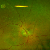
 RAP Lesions
Sep 29 2014 by Thomas A. Ciulla, MD, MBA, FASRS
Fluorescein angiogram of an 81-year-old man revealing several RAP lesions superior to fovea.
Photographer: Stuart Alfred CRA
Condition/keywords: choroidal neovascular membrane (CNVM), neovascular age-related macular degeneration (AMD), retinal angiomatous proliferation (RAP), wet age-related macular degeneration (wet AMD)
-
 ARMD Laser Rx
Aug 7 2013 by H. Michael Lambert, MD
ARMD Laser Rx
Condition/keywords: wet age-related macular degeneration (wet AMD)
-
 Silicone Oil Large and Small Droplets 8 Days Post Avastin (Avella) Injection
Aug 16 2016 by Paul E. Tornambe, MD
The 88-year-old man had an Avastin injection (compounded by Avella) OD for AMD 8 days prior to this photo. He immediately complained of floaters after the injection which persisted 8 days later. The Optos photo, taken 8 days after the injection, shows a large silicone oil bubble about 1DD in size above the ST arcade and more than a dozen smaller 0.1DD bubbles over the macula suspended in the vitreous gel.
Photographer: Louanna Boren, Retina Consultants, San Diego
Condition/keywords: silicone oil, wet age-related macular degeneration (wet AMD)
-
 RPE Tear: Fluorescein Angiography
May 2 2015 by Thomas A. Ciulla, MD, MBA, FASRS
Mid Phase Fluorescein Angiogram: The scrolled and redundant RPE just temporal to the fovea blocks underlying choroidal fluorescence. The absent RPE, more temporally, results in a window defect with intense hyperfluorescence.
Photographer: Stuart Alfred
Condition/keywords: choroidal neovascular membrane (CNVM), retinal pigment epithelium (RPE) tear, wet age-related macular degeneration (wet AMD)
-
 ARMD Laser Rx
Aug 7 2013 by H. Michael Lambert, MD
ARMD Laser Rx
Condition/keywords: wet age-related macular degeneration (wet AMD)
 Loading…