 Initializing download.
-
 By Thomas A. Ciulla, MD, MBA, FASRS
Indiana University School of Medicine - Uploaded on May 2, 2015.
- Last modified by Chayal Patel on May 7, 2015.
- Rating
- Appears in
- RPE Tear
- Condition/keywords
- retinal pigment epithelium (RPE) tear, wet age-related macular degeneration (wet AMD), choroidal neovascular membrane (CNVM)
- Photographer
- Stuart Alfred
- Imaging device
- Scanning laser ophthalmoscope
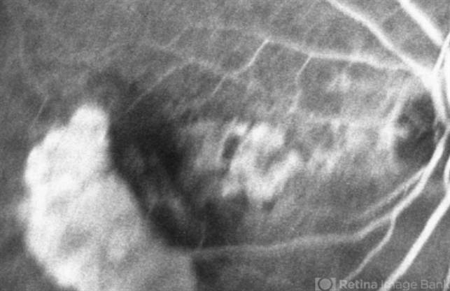
- Description
- Mid Phase Fluorescein Angiogram: The scrolled and redundant RPE just temporal to the fovea blocks underlying choroidal fluorescence. The absent RPE, more temporally, results in a window defect with intense hyperfluorescence.


---thumb.JPG/image-square;max$79,0.ImageHandler "Advanced Wet AMD")