Search results (51 results)
-
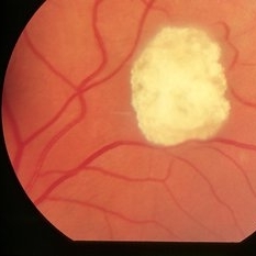
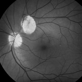
 Astrocytoma
Mar 13 2013 by Jose Dalma-Weiszhausz, MD
28-year-old male with tuberous sclerosis.
Photographer: José Dalma, MD, Dalma & Asoc. Mexico City, Mexico
Condition/keywords: astrocytoma, tuberous sclerosis
-
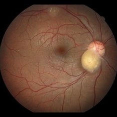
---thumb.jpg/image-square;max$300,300.ImageHandler) Astrocytic Hamartoma
Astrocytic Hamartoma
Feb 20 2013 by From the Collections of Thomas M. Aaberg, MD and Thomas M. Aaberg Jr., MD
Mulberry lesion.
Condition/keywords: tuberous sclerosis
-
 Tuberous Sclerosis
Oct 9 2012 by Alan D. Letson, MD
Small astrocytic hamartoma in asymptomatic 65-year-old woman with Tuberous sclerosis.
Photographer: Beverly Radcliffe
Condition/keywords: astrocytoma, hamartoma, tuberous sclerosis
-
---thumb.jpg/image-square;max$300,300.ImageHandler) Tuberous Sclerosis
Tuberous Sclerosis
Feb 13 2013 by From the Collections of Thomas M. Aaberg, MD and Thomas M. Aaberg Jr., MD
Mulberry lesion.
Condition/keywords: peripapillary, tuberous sclerosis
-
 Astrocytic hamartoma of the retina
Jan 11 2013 by Alex P. Hunyor, MD
Astrocytic hamartoma in a patient with tuberous sclerosis. Note also vascular sheathing inferior to optic disc
Condition/keywords: tuberous sclerosis
-
 Retina Hamartomas in Tuberous Sclerosis
Jan 8 2019 by Sofia Mano
Female 19-years-old with tuberous sclerosis. BCVA LE 10/10. Fundus LE shows three multinodular hamartomas.
Photographer: Sofia Sousa Mano
Imaging device: Canon CR 2 plus
Condition/keywords: hamartoma, tuberculous chorioretinitis
-
 Astrocytoma
Feb 9 2015 by Patricia Araújo
Fundus photography of an 15-year-old boy with tuberous sclerosis.
Photographer: Dr Patricia Correa
Condition/keywords: astrocytic hamartoma, tuberous sclerosis
-
 Astrocytic Hamartoma
Apr 30 2015 by Mariam A Al-Feky, MD
A 15-year-old boy with history of seizures controlled on treatment. C/O: OD painless DV 10/7 ago (accidental discovery) O/E: BCVA OD: 6/60 ,, OS 6/6. AS: NAD OU. Pupil: RRR no RAPD OU. Fundus examination OD showed a retinitis like lesion with an overlying corkscrew vessel well evident on FFA with late leakage and CSR and OCT through the retinitis like lesion shows diffuse hypereflective thickeninig in the superficial NFL. Thorough history taking revealed that patient has seizures and MRI lesions suggestive of tuberous sclerosis. So this is exudative hamartoma secondary to tuberous sclerosis with marked resolution after single IVI of Lucentis. Retinitis like lesion with corkscrew vessels in FFA is typical together with the homogenous hypereflective thickening in the NFL.
Photographer: Mariam AL-Feky
Imaging device: Optical coherence tomography
Condition/keywords: astrocytic hamartoma
-
 Tuberous Sclerosis
Jan 8 2015 by H. Michael Lambert, MD
Large astrocytic hamartoma along IT arcade.
Condition/keywords: tuberous sclerosis
-
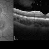
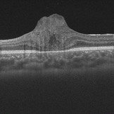
 Astrocytic Hamartoma from Tuberous Sclerosis
Sep 18 2016 by John T. Thompson, MD
OCT of astrocytic hamartoma in child with tuberous sclerosis.
Imaging device: Heidelberg Spectralis
Condition/keywords: astrocytic hamartoma, tuberous sclerosis
-
 Hamartoma Tuberous Sclerosis
Jun 2 2016 by Nelson Chamma Capelanes, MD
Fundus photograph of an 52-year-old man with tuberous sclerosis and retinal hamartoma.
Photographer: Nelson Chamma Capelanes, Fundação Hilton Rocha, Promédica Indaiatuba, Brazil
Condition/keywords: tuberous sclerosis
-
 Multiple Astrocytic Hamartomas
Jul 26 2018 by Olivia Rainey
Optical coherence tomography of a 7-year-old female with multiple astrocytic harmartomas as a retinal manifestation of tuberous sclerosis. Patient came to our office to rule out possible drug toxicity from Sabril, a an anticonvulsant. There were no signs of retinal toxicity by extended ophthalmoscopy or imaging, yet she will be monitored every 6 months.
Photographer: Olivia Rainey
Imaging device: Heidelberg Spectralis
Condition/keywords: astrocytic hamartoma, Heidelburg Spectralis, infrared image, left eye, optical coherence tomography (OCT), tuberous sclerosis
-
 Tuberous Sclerosis
Jan 8 2015 by H. Michael Lambert, MD
Color image of peripapillary astrocytic hamartoma.
Condition/keywords: astrocytic hamartoma, tuberous sclerosis
-
 Retinal Astrocytic Hamartoma
Feb 11 2017 by Alexandra L Pappas, MD, MS
55-year-old male with an incidental finding. No other systemic findings of tuberous sclerosis.
Photographer: Cheri Scully, LPN
Condition/keywords: astrocytic hamartoma
-
 Tuberous Sclerosis
Jan 8 2015 by H. Michael Lambert, MD
Tuberous Sclerosis with peri-ungal fibroma.
Condition/keywords: tuberous sclerosis
-
 Tuberous Sclerosis
Jan 8 2015 by H. Michael Lambert, MD
Astrocytic hamartoma.
Condition/keywords: astrocytic hamartoma, tuberous sclerosis
-
 Tuberous Sclerosis
Jan 8 2015 by H. Michael Lambert, MD
Peripapillary astrocytic hamartoma.
Condition/keywords: tuberous sclerosis
-
 Tuberous Sclerosis
Jan 8 2015 by H. Michael Lambert, MD
Tuberous Sclerosis with hypomelanotic skin lesion.
Condition/keywords: tuberous sclerosis
-
 Tuberous Sclerosis
Jan 8 2015 by H. Michael Lambert, MD
Full face view of patient with adenoma sebaceum.
Condition/keywords: tuberous sclerosis
-
 Tuberous Sclerosis
Jan 8 2015 by H. Michael Lambert, MD
Close up image of nasal adenoma sebaceum.
Condition/keywords: tuberous sclerosis
-
 Tuberous Sclerosis
Jan 8 2015 by H. Michael Lambert, MD
Histopathology of astrocytic hamartoma.
Condition/keywords: tuberous sclerosis
-
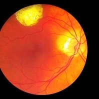
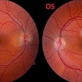 Retinal Phakomatous Hamartoma in Tuberous Sclerosis- Fundus Photos
Aug 28 2019 by Nisarg Joshi, MD
26-year-old female with history of tuberous sclerosis was found to have retinal phakomatous hamartomas in the right eye (peripapillary pale yellow lesion) and the left eye (opaque lesion in superior macula). The macula also shows RPE changes in both eyes.
Photographer: Nisarg Joshi, Geisinger Eye Institute, Danville, PA
Condition/keywords: hamartoma, phakoma, tuberous sclerosis
-
 Tuberous Sclerosis
Jan 8 2015 by H. Michael Lambert, MD
Tuberous Sclerosis- skull X-ray showing intracranial calcification.
Condition/keywords: tuberous sclerosis
-
 Astrocytoma OCT
Jan 9 2018 by Sidra Zafar
Swept source OCT imaging of retinal astrocytoma in a female child with known diagnosis of tuberous sclerosis.
Imaging device: Swept Source
Condition/keywords: astrocytoma, optical coherence tomography (OCT), tuberous sclerosis
-
 Tuberous Sclerosis
Jan 8 2015 by H. Michael Lambert, MD
Color image of peripapillary astrocytic hamartoma.
Condition/keywords: tuberous sclerosis
 Loading…