Search results (90 results)
-
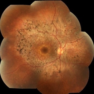
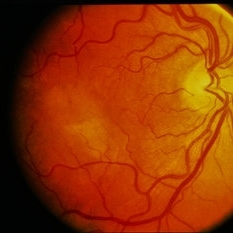
 Acute Syphilitic Posterior Placoid Chorioretinitis
Aug 23 2012 by Gerardo Garcia-Aguirre, MD
Fundus photograph of a 42 year-old male with positive VDRL and FTA-ABS, with a yellowish placoid lesion in the posterior pole.
Photographer: Ricardo Montoya, Asociación para Evitar la Ceguera en México
Condition/keywords: acute syphilitic posterior placoid chorioretinitis, syphilis
-
 Syphilitic Panuveitis, Left Eye
Oct 8 2012 by Pauline T Merrill, MD, FASRS
Left fundus photograph of a 23-year-old Hispanic male with decreased vision for 1 month, recent pain/redness both eyes. Found to have panuveitis; also rash on palms & soles. Labs positive for FTA-ABS, and CSR VDRL. Treated with IV penicillin for 14 days.
Photographer: Karen Parque, Illinois Retina Associates, Chicago, IL
Condition/keywords: panuveitis, snowballs, syphilis, vitritis
-
 Syphilis Neuroretinopathy
Apr 2 2018 by JEFFERSON R SOUSA, Tecg.º (Biomedical Systems Technology)
Female patient, 21-years-old, with complaint of low vision in the right eye for 3 years. According to information from the patient's history, at the time she noticed the low vision, it also coincided with a picture of a strong urinary infection as well as episodes of constant tonsillitis. Yes, the patient did not seek medical attention and self-medicated with antibiotics. In ophthalmologic evaluation, as well as examinations of color retinography and ocular fundus autofluorescence, important pigmentary alterations were observed following vascular arches with pigment mobilization in osteoclasts (aspect of a unilateral pigmentary retinitis secondary to the inflammatory process). Which suggested inflammatory process sequelae. Through the laboratory tests, he had positive (+) confirmation for SYPHILIS NEURORETINOPATHY .
Photographer: JEFFERSON R SOUSA - Study Center and Ophthalmological Research Dr. Andre M V Gomes, Institute Dr. Suel Abujamra São Paulo-Brazil
Imaging device: Fundus camera Topcon TRC-50 DX, Imaginet 5.0, angle de 50 graus. Flash 36 / Mosaic with 10 images.
Condition/keywords: neurosyphilitic optic atrophy, retinitis pigmentosa, syphilis, syphilis neuroretinopathy
-
 Syphilitic Panuveitis, Right Eye
Oct 8 2012 by Pauline T Merrill, MD, FASRS
Right fundus photograph of a 23-year-old Hispanic male with decreased vision for 1 month, recent pain/redness both eyes. Found to have panuveitis; also rash on palms & soles. Labs positive for FTA-ABS, and CSR VDRL. Treated with IV penicillin for 14 days.
Photographer: Karen Parque, Illinois Retina Associates, Chicago, IL
Condition/keywords: panuveitis, snowballs, syphilis, vitritis
-
 Acute Syphilitic Posterior Placoid Chorioretinitis
Aug 23 2012 by Gerardo Garcia-Aguirre, MD
Early phase fluorescein angiogram of a 42 year-old male, showing hyperflourescence with a granular pattern in the posterior pole.
Photographer: Ricardo Montoya, Asociación para Evitar la Ceguera en México
Condition/keywords: acute syphilitic posterior placoid chorioretinitis, syphilis
-
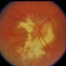
 Syphyllis
Aug 15 2013 by From the Collections of Thomas M. Aaberg, MD and Thomas M. Aaberg Jr., MD
Palmar rash.
Condition/keywords: syphilis
-
---thumb.jpg/image-square;max$300,300.ImageHandler) Syphyllis
Syphyllis
Aug 15 2013 by From the Collections of Thomas M. Aaberg, MD and Thomas M. Aaberg Jr., MD
Acute retinitis.
Condition/keywords: syphilis
-
 Acute Syphilitic Posterior Placoid Chorioretinitis
Aug 23 2012 by Gerardo Garcia-Aguirre, MD
Fluorescein angiogram of a 42 year-old male, showing hyperflourescence with a granular pattern in the posterior pole.
Photographer: Ricardo Montoya, Asociación para Evitar la Ceguera en México
Condition/keywords: acute syphilitic posterior placoid chorioretinitis, syphilis
-
 Syphyllis
Aug 15 2013 by From the Collections of Thomas M. Aaberg, MD and Thomas M. Aaberg Jr., MD
Acute retinitis.
Condition/keywords: syphilis
-
 Congenital Syphilis
Feb 20 2015 by H. Michael Lambert, MD
Color photo of chorioretinitis.
Condition/keywords: color photo, congenital, syphilis
-
 Eales Disease
Apr 26 2013 by Howard Schatz, MD
Eales b/c Behcet's syphilis.
Condition/keywords: Eales disease, syphilis
-
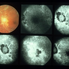
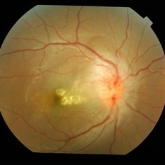
 Syphilis Neuroretinopathy (Color Photo)
Sep 25 2013 by Alexandre Durao Alves Pereira, MD
Syphilis neuroretinopathy, late phase FA, (color photograph).
Photographer: Alexandre Pereira
Condition/keywords: color photo, late phase, syphilis neuroretinopathy
-
 Syphilitic Chorioretinitis
Apr 8 2019 by Gary R. Cook, MD, FACS
68-year-old patient with 'salt and pepper'-type diffuse chorioretinitis OD secondary to acquired syphilis; V.A. = 20/25
Imaging device: Topcon VT-50
Condition/keywords: chorioretinitis, pseudo retinitis pigmentosa, syphilis
-
 Syphilis Neuroretinopathy FA
Sep 25 2013 by Alexandre Durao Alves Pereira, MD
Syphilis neuroretinopathy, late phase FA.
Photographer: Alexandre Pereira
Condition/keywords: FA late phase
-
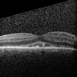 Syphilis Pre Treatment OCT
Sep 1 2017 by Annal D Meleth, MD, MS
Syphilis pre treatment OCT.
Photographer: Kenneth Thompson
Condition/keywords: acute syphilitic posterior placoid chorioretinitis, syphilis
-
 Syphyllis
Aug 15 2013 by From the Collections of Thomas M. Aaberg, MD and Thomas M. Aaberg Jr., MD
Acute retinitis.
Condition/keywords: syphilis
-
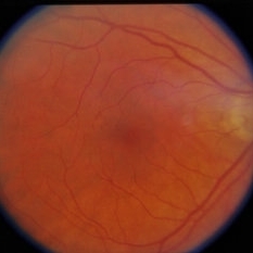
 Syphilis Placoid Epitheliopathy
May 15 2014 by David Callanan, MD
50-year-old patient, syphilis placoid epitheliopathy.
Condition/keywords: placoid, syphilis
-
 Syphyllis
Aug 15 2013 by From the Collections of Thomas M. Aaberg, MD and Thomas M. Aaberg Jr., MD
Acute retinitis.
Condition/keywords: syphilis
-
 Congenital Syphilis
Feb 20 2015 by H. Michael Lambert, MD
Color photo of chorioretinitis.
Condition/keywords: color photo, congenital, syphilis
-
 Neuroretinitis
Apr 19 2014 by Mallika Goyal, MD
Right eye fundus of a 25-year-old female patient with idiopathic neuroretinitis shows disc edema and congestion with macular exudates and edema at presentation. All relevant investigations (to rule out sarcoidosis, TB, SLE, leptospirosis, syphilis, borrelia bergdorferi, HIV returned negative). She was treated with pulsed intravenous steroids with resolution.
Photographer: Mallika Goyal, MD, Apollo Health City, Hyderabad, India
Condition/keywords: neuroretinitis
-
 Syphilis Neuroretinopathy
Sep 25 2013 by Alexandre Durao Alves Pereira, MD
Syphilis neuroretinopathy late phase FA.
Photographer: Alexandre Pereira
Condition/keywords: late phase
-
 Syphilis Neuroretinopathy (Red Free Photo)
Sep 25 2013 by Alexandre Durao Alves Pereira, MD
Syphilis neuroretinopathy, late phase FA, (red free photograph).
Photographer: Alexandre Pereira
Condition/keywords: late phase, red-free
-
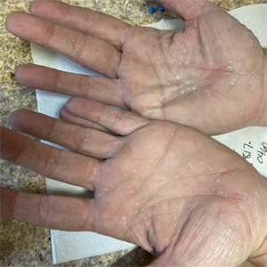 Palms of Patient with Placoid Lesions in Posterior Segment
Dec 20 2020 by John S. King, MD
44-year-old white female seen over the weekend complaining of a "spot" in her vision centrally OD for three days. She was referred over by another eye doctor who was concerned about a possible retinal detachment vs ARN in that eye. Her past medical history includes adrenal insufficiency for which she takes a low dose of hydrocortisone, thyroxine (post thyroidectomy), Plaquenil (inflammatory arthritis). She is divorced with one partner and denies any IVDU. Va 20/200 OD and 20/20 OS, IOP 12 OU, Pupils mydriatic post gtts (light desaturation OD). There was 1+ A/C cell OD, O/W unremarkable anterior segment OU; in the posterior segment OD there was 1+ vitritis with a diffusely swollen optic disc and a large yellowish placoid lesion in the macula with yellowish border and extended out past the arcades inferiorly, as well as another lesion smaller in the IN periphery. There was trace vitreous cell OS, mild disc edema, and a large, granular placoid area nasally that appeared to be granulated. The OCT showed mild subfoveal fluid with nodular areas in the RPE and some overlying irregular architecture of the outer retina. Syphilis was a concern at this point. She denied any hand or foot rash, and said that she was recently working on the house, and her hands were dried out. There did appear to be a rash on the hand (See Image), and later learned that she had a rash on the soles of her feet. She was sent to ED for a work-up and her syphilis IgG was positive and VDRL 1:128, and negative for HIV. She was started on a course IV Penicillin (40mg PO steroid two days after tx started). She has responded well. A few days after treatment her visual acuity has improved to 20/60 OD; there was no anterior segment inflammation OU, and decreased vitreous cell OU. Disc edema was improved. The large placoid lesion in the macula of the right eye was slightly enlarged, but more granular in appearance without a distinct yellowish border, and the smaller lesions SN had dissipated. OCT showed resolution of the subfoveal fluid and an improved appearance of the outer retina and RPE layer.
Imaging device: Optos CA
Condition/keywords: acute syphilitic posterior placoid chorioretinitis
-
 Syphilis Placoid Epitheliopathy
May 15 2014 by David Callanan, MD
50-year-old patient, syphilis placoid epitheliopathy.
Condition/keywords: placoid, syphilis
-
 Syphilis Neuroretinopathy (Late Phase FA)
Sep 25 2013 by Alexandre Durao Alves Pereira, MD
Syphilis neuroretinopathy, (late phase FA).
Photographer: Alexandre Pereira
Condition/keywords: late phase
 Loading…