Search results (43 results)
-
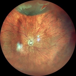
 Acute Superior Retinal Detachment
Oct 12 2012 by Jeffrey G. Gross, MD, FASRS
Acute, superior retinal detachment.
Condition/keywords: acute
-
 Cytomegalovirus Retinitis, Healed
Sep 27 2012 by Jeffrey G. Gross, MD, FASRS
CMV retinitis, healed, superior retina.
Condition/keywords: healed, retinitis, superior retina
-
 Choroidal Melanoma
Feb 2 2018 by Olivia Rainey
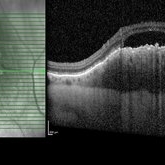
Optical coherence tomography with enhanced depth imaging of a 78-year-old female with choroidal melanoma with subretinal fluid affecting her right eye.
Photographer: Olivia Rainey
Imaging device: Heidelberg Spectralis
Condition/keywords: enhanced depth imaging, infrared image, optical coherence tomography (OCT), subretinal fluid, superior retina
-
 Choroidal metastasis
May 2 2013 by Henry J. Kaplan, MD
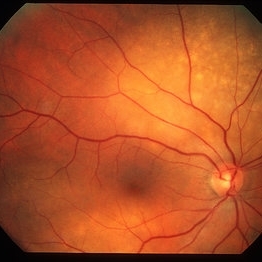
Choroidal metastasis from breast cancer in the superior retina as a yellow lesion with Peau d'Orange appearance. Right eye.
Condition/keywords: choroidal metastasis
-
 Valsalva retinopathy severe
Oct 22 2012 by Ronald C. Gentile, MD
A 46-year-old women noticed inferior field loss just inferior to fixation in her right eye following a bout of food poisoning and intense vomiting. A sub-hyaloid hemorrhage was noted to be layering just superior to the center of the fovea that appeared to have originated from the superior retinal arcade.
Photographer: The New York Eye & Ear Infirmary Department of Medical Imaging
Condition/keywords: valsalva retinopathy
-
 Acute Superior Retinal Detachment
Oct 12 2012 by Jeffrey G. Gross, MD, FASRS
Acute superior RD.
-
 Asymptomatic Superior Retinal Detachment
May 5 2016 by Steven J Ryder, MD
38-year-old African American female with moderate myopia (-4.50 Sph OU) and asymptomatic superior retinal detachment in the right eye. Montage fundus photography showing localized retinal detachment superiorly with single full-thickness retinal break at 12:00.
Photographer: Luis Bernhard, Miami VA Healthcare System
Imaging device: Topcon
Condition/keywords: asymptomatic, full thickness retinal hole, myopia, retinal break, retinal detachment with retinal defect
-
 RD with Giant Tear After Trauma
Jan 1 2013 by John T. Thompson, MD
Retinal detachment with giant retinal tear after blunt trauma, superior retina is folded over itself.
Condition/keywords: blunt trauma, retinal tear
-
 Superior Bullous Detachment With Horseshoe Tear
May 15 2014 by Manish Nagpal, MD, FRCS (UK), FASRS
Patient having bullous superior retinal detachment with a horseshoe tear.
Photographer: pooja barot, Optometrist, Retina Foundation, Ahmedabad
-
---thumb.JPG/image-square;max$300,300.ImageHandler) Superior Retinal Detachment.
Superior Retinal Detachment.
Jul 10 2013 by Jason S. Calhoun
Fundus photograph shows small retinal detachment at 12-o'clock superiorly in the right eye.
Photographer: Jason S. Calhoun, Department of Ophthalmology, Mayo Clinic Jacksonville, Florida
-
 Sectoral Retinitis Pigmentosa
May 4 2015 by Mallika Goyal, MD
Left fundus of a 46-year-old lady with retinitis pigmentosa affecting inferior retinal quadrants; superior retina, optic nerve head and macula are normal. She is asymptomatic and fundus picture has been stable over 10 years follow-up. She is the offspring of a consanguineous marriage.
Photographer: Mallika Goyal, MD, Apollo Health City, Jubilee Hills, Hyderabad
Condition/keywords: retinal pigmentosa
-
 Bullous Retinoschisis with Outer Retinal Holes
Jun 15 2020 by Olivia Rainey
Ultra-widefield pseudocolor fundus photograph of a 56-year-old female with bullous retinoschisis with outer retinal holes affecting her right eye. The physician noted superotemporal retinoschisis in her monoculcar functioning eye. There was no demarcation line and no inner or outer layer breaks at her first appointment in February of 2020. On 6/15/20 she had a new onset outer holes and SRF tracking inferiorly. The physician recommended observation, however if this continues to progress we have discussed indications for barrier laser.
Photographer: Olivia Rainey, OCT-C, COA
Imaging device: Optos California
Condition/keywords: bullous retinoschisis, Optos, outer layer breaks, outer layer hole, pseudocolor, subretinal fluid, superior retina, ultra-wide field imaging
-
 Sectoral Retinitis Pigmentosa
May 4 2015 by Mallika Goyal, MD
Right fundus of a 46-year-old lady with retinitis pigmentosa affecting inferior retinal quadrants; superior retina, optic nerve head and macula are normal. She is asymptomatic and fundus picture has been stable over 10 years follow-up. She is the offspring of a consanguineous marriage.
Photographer: Mallika Goyal, MD, Apollo Health City, Jubilee Hills, Hyderabad
Condition/keywords: retinal pigmentosa
-
 Asymptomatic Superior Retinal Detachment
May 5 2016 by Steven J Ryder, MD
38-year-old African American female with moderate myopia (-4.50 Sph OU) and asymptomatic superior retinal detachment in the right eye. Zeiss Cirrus OCT capturing full-thickness retinal break at 12:00 and temporal vitreoretinal traction.
Photographer: Luis Bernhard, Miami VA Healthcare System
Imaging device: Zeiss Cirrus
Condition/keywords: asymptomatic, full thickness retinal hole, retinal break, retinal detachment with retinal defect
-
 Sectoral Retinitis Pigmentosa
May 4 2015 by Mallika Goyal, MD
Right fundus of a 46-year-old lady with retinitis pigmentosa affecting inferior retinal quadrants; superior retina, optic nerve head and macula are normal. She is asymptomatic and fundus picture has been stable over 10 years follow-up. She is the offspring of a consanguineous marriage.
Photographer: Mallika Goyal, MD, Apollo Health City, Jubilee Hills, Hyderabad
Condition/keywords: retinal pigmentosa
-
 Sectoral Retinitis Pigmentosa
May 4 2015 by Mallika Goyal, MD
Left fundus of a 46-year-old lady with retinitis pigmentosa affecting inferior retinal quadrants; superior retina, optic nerve head and macula are normal. She is asymptomatic and fundus picture has been stable over 10 years follow-up. She is the offspring of a consanguineous marriage.
Photographer: Mallika Goyal, MD, Apollo Health City, Jubilee Hills, Hyderabad
Condition/keywords: retinal pigmentosa
-
 Asymptomatic Superior Retinal Detachment
May 5 2016 by Steven J Ryder, MD
38-year-old African American female with moderate myopia (-4.50 Sph OU) and asymptomatic superior retinal detachment in the right eye. Zeiss OCT capturing vertical raster scans through border of retinal detachment.
Photographer: Luis Bernhard, Miami VA Healthcare System
Imaging device: Cirrus
Condition/keywords: asymptomatic, full thickness retinal hole, retinal detachment with retinal defect
-
 Sectoral Retinitis Pigmentosa
May 4 2015 by Mallika Goyal, MD
Left fundus of a 46-year-old lady with retinitis pigmentosa affecting inferior retinal quadrants; superior retina, optic nerve head and macula are normal. She is asymptomatic and fundus picture has been stable over 10 years follow-up. She is the offspring of a consanguineous marriage.
Photographer: Mallika Goyal, MD, Apollo Health City, Jubilee Hills, Hyderabad
Condition/keywords: retinal pigmentosa
-
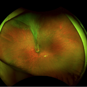
 Total retinal Detachment multiple holes
Sep 26 2022 by Denica Rodriguez
60 year old Male presented with two week old Macula off Retinal detachment with multiple tears.
Photographer: Denica Rodriguez
Imaging device: Optos California
Condition/keywords: color fundus photograph, color photo, macula-off, optos, pseudocolor, Retinal detachment, retinal holes, retinal tear, Retinal tear with detachment, superior arcade, superior field, superior retina, total retinal detachment
-
 Choroidal Detachment
Jan 17 2022 by Logan ryzenga
Left ultra-wide field photograph of an 81-year old female with a choroidal detachment affecting her left eye. Patient had a stent placed November, 2021 and following the procedure she complains of variable blurred vision and severe constricted visual fields. She presented at our office with flashes a month prior but without pain or floaters.
Photographer: Logan Ryzenga
Imaging device: Optos California
Condition/keywords: choroidal detachment, fundus photograph, left eye, Optos, pseudocolor, superior retina, ultra-wide field imaging
-
 Fresh Superior Macula-On Rhegmatogenous Retinal Detachment
Feb 10 2018 by Deepak Bhojwani, MS
A 60 year old gentlemen came rushing to the retinal clinic with history of sudden onset of loss of inferior visual field since last 3 hours. Fundus photograph indeed corelates with his complaints documenting fresh superior macula-on rhegmatogenous retinal detachment.
Photographer: Dr Deepak Bhojwani, Raghudeep Eye Hospital , Ahmedabad
Condition/keywords: macula-on fresh superior retinal detachment
-
 Retinal Detachment with Horseshoe Tear
Jun 10 2020 by Manish Nagpal, MD, FRCS (UK), FASRS
Localized superior retinal detachment with horseshoe tear and minimal fluid.
Photographer: gayathri mohan
Imaging device: nidek slo mirante
-
 Ocular Toxocariasis with Peripheral Granuloma
Apr 24 2021 by Alexandre Grandinetti, MD, PhD
8-year-old boy with a retinal fold secondary to peripheral toxocara canis granuloma localized on the superior retina.
Photographer: Corina Skrzek
Imaging device: Optos California
Condition/keywords: toxocariasis
-
 Retinal Detachment with Retinal Tears
Dec 11 2018 by Olivia Rainey
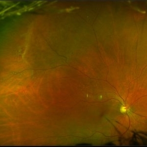
Ultra-wide field pseudocolor image of a 56-year-old male with a large superior retinal detachment with retinal tears affecting his right eye.
Photographer: Olivia Rainey
Imaging device: Optos
Condition/keywords: Optos, pseudocolor, retinal tear with detachment
-
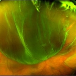
 Traumatic Giant Retinal Tear Associated Retinal Detachment
Nov 9 2019 by Luis J Haddock, MD
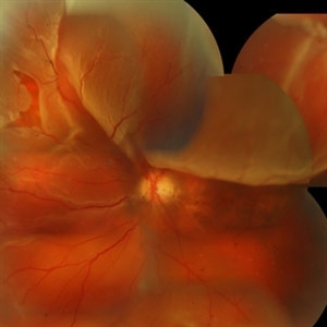
This wide field fundus photograph of the left eye shows a traumatic giant retinal tear associated with total retinal detachment. The image shows the torn superior retina folded over the macula with the underside of the retina visible. There is associated peripheral choroidal detachment due to hypotony from giant retinal tear. This patient has history of spondyloepithelial dysplasia with dwarfism and presented with vision loss after a recent blunt trauma with elbow to the eye.
Imaging device: Optos
Condition/keywords: giant retinal tear, traumatic optic neuropathy
 Loading…