Search results (453 results)
-
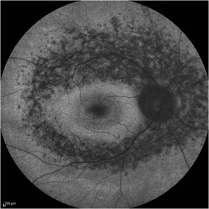 Retinitis Pigmentosa - Fundus Autofluorescence
Sep 20 2014 by Rameez N Hussain, MD
Fundus autofluorescence of retinitis pigmentosa showing hyperautofluorescent rings or foveal hyperautofluorescence.
Photographer: Dr.Rameez N Hussain, MD, Central Imaging Center, Vitreo Retinal Services, Giridhar Eye Institute, Cochin, India
Imaging device: Heidelberg Blue Peak Autofluorescence imaging.
Condition/keywords: bone spicule, cystoid macular edema (CME), fundus autofluorescence (FAF), retinitis pigmentosa
-
 Sector Retinitis Pigmentosa
Oct 8 2012 by Jeffrey G. Gross, MD, FASRS
Sector retinitis pigmentosa, 20/20, left eye.
Condition/keywords: 20/20, left eye, sector retinitis pigmentosa
-
 Retinitis Pigmentosa
Sep 11 2012 by Hamid Ahmadieh, MD
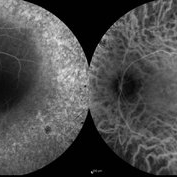
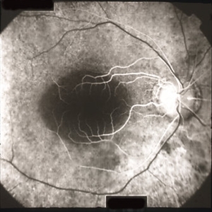
FA & ICG angiography images of a 40-year-old man with RP.
Photographer: Hamid Ahmadieh, MD, Ophthalmic Research Center, Labbafinejad Medical Center, Shahid Beheshti University of Medical Sciences
Imaging device: Heidelberg Spectralis
Condition/keywords: indocyanine green (ICG) angiography, retinitis pigmentosa
-
 Retinitis Pigmentosa
Apr 6 2022 by Marianna Kavalaraki, MD, Msc
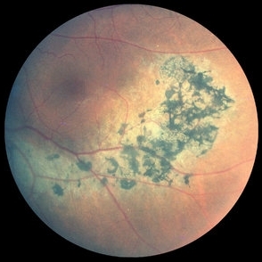
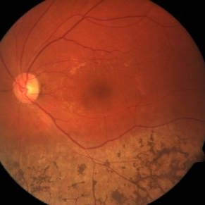
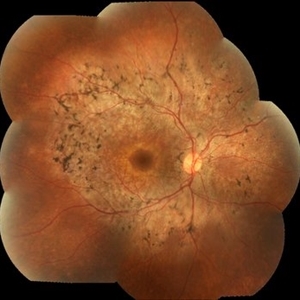
Fundus photography of a 21-year-old man with retinitis pigmentosa. Fundus findings include retinal pigmentary changes in the form of widespread pigment clumpings predominantly in the mid-peripheral fundus, arteriolar attenuation, RPE and retinal atrophy in the posterior pole.
Photographer: Marianna Kavalaraki, General Hospital of Nikaia Piraeus, Department of Ophthalmology
Imaging device: Canon CF-60DSi Digital Fundus Camera
Condition/keywords: retinitis pigmentosa
-
 Sector Retinitis Pigmentosa
Mar 13 2014 by Hyung-Woo Kwak, MD
Fundus photograph of an 57-year-old woman with a sector retinitis pigmentosa. Regionalized areas of bone spicule pigmentation is in the inferior quadrants of the retina.
Photographer: Missok Lee, Kyung Hee University Hospital, Seoul, Korea
Imaging device: Zeiss F450 Plus
Condition/keywords: sector retinitis pigmentosa
-
 Retinitis Pigmentosa
May 26 2017 by Olivia Rainey
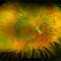
Ultra-wide-field pseudocolor image of the right eye of an 39-year-old female with Retinitis Pigmentosa. She had slightly atypical appearance due to asymmetry: sectoral atrophy in left eye, compared to 360 degree bone spicule formation in right eye. Ddx: Pigmentary degeneration vs infection vs X-linked RP carrier due to asymmetry. Recommended genetic testing through My Retina Tracker, as well as visual field and ERG testing. Patient's vision was sc20/100 PH 20/70 in the right eye and sc20/80 PH 20/40 in the left.
Photographer: Olivia Rainey
Imaging device: Optos California
Condition/keywords: bone spicule, fundus photograph, Optos, peripheral bone spicules, pseudocolor, retinitis pigmentosa, ultra-wide field imaging
-
 Sector Retinitis Pigmentosa
Oct 8 2012 by Jeffrey G. Gross, MD, FASRS
Sector retinitis pigmentosa, 20/20, right eye.
Condition/keywords: 20/20, sector retinitis pigmentosa
-
 Retinitis Pigmentosa
Sep 2 2012 by Hyung-Woo Kwak, MD
A mild pigment epithelial atrophy in the mid-periphery with small white dots at the level of the RPE in fundus.
Imaging device: Zeiss F450 plus
Condition/keywords: retinitis pigmentosa
-
---thumb.jpg/image-square;max$300,300.ImageHandler) Retinitis Pigmentosa and Cataract
Retinitis Pigmentosa and Cataract
Dec 27 2013 by David Callanan, MD
23-year-old male patient, 20/100 OU.
Condition/keywords: cataract, retinitis pigmentosa
-
 Pericentral Retinitis Pigmentosa
Oct 19 2012 by Larry Halperin, MD
Pericentral retinitis pigmentosa
Condition/keywords: pericentral retinitis pigmentosa
-
 Retinitis Pigmentosa - Autofluorescence OD
Jun 18 2018 by Hosam Attia, MD
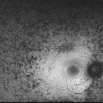
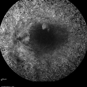
Ultra-wide fundus auto-fluorescence photograph of a 38-year-old African, American female with degenerative myopia, unilateral RP variant, depicting extensive mid-peripheral bone spicules hypo-autofluorescence, extending further into the periphery w/ relative sparing of the macula OD VF 30-V showed severe peripheral constriction OD, enlarged BS OS and OCT showed severe ellipsoid zone degeneration with saucerization and cystoid macular degeneration with no obvious late macular leakage on FA (Both, not shown)
Imaging device: Optos California
Condition/keywords: bone spicule, peripheral bone spicules, retinitis pigmentosa
-
---thumb.jpg/image-square;max$300,300.ImageHandler) Retinitis Pigmentosa
Retinitis Pigmentosa
Oct 13 2012 by Geoffrey G. Emerson, MD, PhD, FASRS
Condition/keywords: bone spicule, retinitis pigmentosa
-
 Retinitis Pigmentosa
Aug 23 2012 by Gerardo Garcia-Aguirre, MD
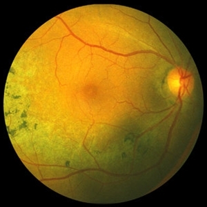
Fundus photograph showing diffuse pigmentary changes with relative sparing of the macula.
Photographer: Noemí Hernández, Asociación para Evitar la Ceguera en México
Imaging device: Zeiss FF4
Condition/keywords: retinitis pigmentosa
-
 Retinitis pigmentosa AD Slide 1
Oct 22 2012 by Ronald C. Gentile, MD
37 year-old man presents with nyctalopia and tunnel vision. Fundus photo reveals sparing of the fovea with accentuation of the foveal thickness compared to the surrounding atrophic retina. There is waxy pallor of the optic nerve and attenuation of the retinal vessels.
Photographer: The New York Eye & Ear Infirmary Department of Medical Imaging
Condition/keywords: retinitis pigmentosa
-
 Retinitis pigmentosa AR slide 1
Oct 22 2012 by Ronald C. Gentile, MD
17 year-old girl, born of consanguineous parents, presented with nyctalopia and poor vision in both eyes. Vision was hand motions in both eyes. Electroretinogram was extinguished for both photopic and scotopic responses.
Photographer: The New York Eye & Ear Infirmary Department of Medical Imaging
Condition/keywords: retinitis pigmentosa
-
 Retinitis pigmentosa AD Slide 4
Oct 22 2012 by Ronald C. Gentile, MD
Early fluorescein angiography revealed early hyper-fluorescence surrounding the fovea consistent with retinal pigment epithelial (RPE) depigmentation. This accentuated the contrast between the normal blockage (hypo-fluorescence) of the macular luteal pigment from the surrounding RPE window defect (hyper-fluorescence).
Photographer: The New York Eye & Ear Infirmary Department of Medical Imaging
Condition/keywords: retinitis pigmentosa
-
 Retinitis Pigmentosa With Hemangioma CF
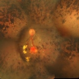
Dec 15 2016 by Manish Nagpal, MD, FRCS (UK), FASRS
Fluorescein angiography OS of a patient having retinitis pigmentosa with a hemangioma inferiorly.
Condition/keywords: hemangioma, retinitis pigmentosa
-
 Retinitis Pigmentosa
Oct 17 2014 by Avris Romario Diparaja Siahaan
A fundus fluorescein angiography of a 25-year-old woman with retinitis pigmentosa in both of her eyes.
Photographer: Renjer Daniel Roring, Klinik Mata Nusantara
Imaging device: Heidelberg Spectralis
Condition/keywords: retinitis pigmentosa, ultra-wide field imaging
-
 Bone Corpuscle Pigments
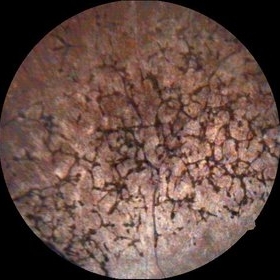
Sep 11 2014 by Mehul A Shah
A 42-year-old female presented with gradual reduction in vision.
Photographer: Drashti Netralaya,Dahod
Imaging device: FF 450
Condition/keywords: retinitis pigmentosa (RP) dystrophy
-
 Retinitis pigmentosa AD Slide 3
Oct 22 2012 by Ronald C. Gentile, MD
Fundus changes were symmetrical. Visual fields revealed small central islands corresponding the the centrally sparred areas of maculae in both eyes.
Photographer: The New York Eye & Ear Infirmary Department of Medical Imaging
Condition/keywords: retinitis pigmentosa
-
 Syphilis Neuroretinopathy
Apr 2 2018 by JEFFERSON R SOUSA, Tecg.º (Biomedical Systems Technology)
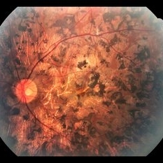
Female patient, 21-years-old, with complaint of low vision in the right eye for 3 years. According to information from the patient's history, at the time she noticed the low vision, it also coincided with a picture of a strong urinary infection as well as episodes of constant tonsillitis. Yes, the patient did not seek medical attention and self-medicated with antibiotics. In ophthalmologic evaluation, as well as examinations of color retinography and ocular fundus autofluorescence, important pigmentary alterations were observed following vascular arches with pigment mobilization in osteoclasts (aspect of a unilateral pigmentary retinitis secondary to the inflammatory process). Which suggested inflammatory process sequelae. Through the laboratory tests, he had positive (+) confirmation for SYPHILIS NEURORETINOPATHY .
Photographer: JEFFERSON R SOUSA - Study Center and Ophthalmological Research Dr. Andre M V Gomes, Institute Dr. Suel Abujamra São Paulo-Brazil
Imaging device: Fundus camera Topcon TRC-50 DX, Imaginet 5.0, angle de 50 graus. Flash 36 / Mosaic with 10 images.
Condition/keywords: neurosyphilitic optic atrophy, retinitis pigmentosa, syphilis, syphilis neuroretinopathy
-
 Macula Spared Advanced Retinitis Pigmentosa 11
Apr 11 2013 by Raj K. Maturi, MD
78-year-old male with advanced retinitis pigmentosa.
Photographer: Tom Steele, CRA Midwesteye Institute, Indianapolis Indiana
Condition/keywords: macula, retinitis pigmentosa
-
 Unilateral Retinitis Pigmentosa
May 1 2014 by Raj K. Maturi, MD
53-year-old woman with significant salt and pepper retinopathy OS.
Photographer: Tom Steele, Midwest Eye Institute
Condition/keywords: retinitis pigmentosa (RP) dystrophy
-
 Retinitis pigmentosa AD Slide 5
Oct 22 2012 by Ronald C. Gentile, MD
Late fluorescein angiography revealed some fading of the early hyper-fluorescence surrounding the fovea consistent with retinal pigment epithelial depigmentation without any late leakage or CME.
Photographer: The New York Eye & Ear Infirmary Department of Medical Imaging
Condition/keywords: retinitis pigmentosa
-
 Autofluorescence of Retinitis Pigmentosa
Jul 13 2016 by Linda A Cernichiaro- Espinosa, MD
Fundus autofluorescence of an 53-year-old woman with retinitis pigmentosa.
Photographer: Tec Ricardo Montoya, Clínica Oftalmológica Anzures
Condition/keywords: retinitis pigmentosa
 Loading…