Search results (101 results)
-
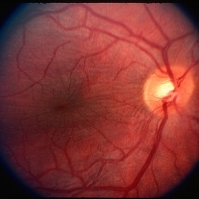 Retinal Folds
Mar 29 2013 by Henry J. Kaplan, MD
Retinal folds as fine wrinklings.
Condition/keywords: retinal fold
-
 Chorioretinal Folds
-
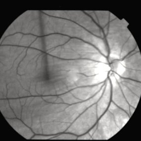
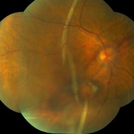 Posterior Retinal Folds
Feb 9 2015 by Leandro C. Zacharias, MD, PhD
Fundus photograph of a 59-year-old woman 3 weeks after buckle for a macula-off retinal detachment.
Photographer: Leandro Cabral Zacharias
Imaging device: Zeiss Visucam
Condition/keywords: retinal fold
-
 Ocular Hypotony Due to Leaking Bleb
Apr 1 2019 by Anfisa Ayalon, MD
81-year-old male who had trabeculectomy in his right eye 4 years ago, presented to the emergency room with complains of decreased vision in that eye for two months. Slit-lamp examination showed cystic bleb with leakage, intraocular pressure was 0 MMHg. Fundus examination showed hypotony maculopathy, peripheral choroidal detachments, multiple chorioretinal folds with subretinal fluid.
Photographer: Anfisa Ayalon, MD., Meir Medical Center, Kfar Saba, Israel.
Imaging device: California, Optos 200 DTX
Condition/keywords: choroidal detachment, hypotonous retinopathy, hypotony maculopathy
-
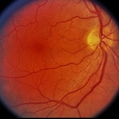
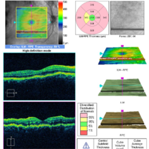
 Choroidal Folds
Nov 28 2014 by Thomas A. Ciulla, MD, MBA, FASRS
This 53-year-old man was noted to have choroidal folds right greater than left. The visual acuity was normal at 20/15. The choroidal folds are visible on OCT, especially on the vertical cuts that image across the horizontal folds. Angiography revealed staining of the folds without CNVM, choroidal mass, or optic nerve edema.
Photographer: Charlotte Harris
Condition/keywords: bilateral chorioretinal folds, choroidal folds
-
 Chorioretinal Fold
Sep 2 2012 by Hyung-Woo Kwak, MD
Chorioretinal folds are seen as coarse striations of the fovea surface after trauma.
Imaging device: Zeiss F450 plus
Condition/keywords: chorioretinal fold
-
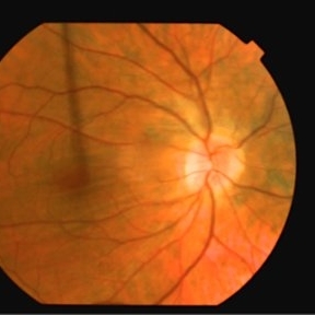
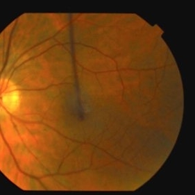
 Retinal Folds After Surgery
Jun 23 2016 by Andrea Arriola-Lopez, MD MSc
45-year-old man with history of rhegmatogenous retinal detachment and segmental scleral buckle from MIX to MXII, SF6 and cryotherapy on right eye was performed. Radial folds on indentation was seen after surgery. Three weeks later, inferior macular folds was found. The patient was asymptomatic. Observation was decided. Retina remains attach. On top, close up to macular area shows inferior folds far from fovea. Bottom, red free photograph shows no RPE changes on the same retina fold area.
Photographer: Andrea E. Arriola-López MD MSc
Imaging device: OPTOS
Condition/keywords: macular fold, retina surgery, scleral buckle
-
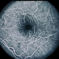
 Retinal Folds Following Retinal Reattachment Surgery
Nov 22 2015 by Mallika Goyal, MD
Multiple retinal folds 4 weeks following vitreous surgery (perfluorodecalin assisted) for retinal detachment with giant retinal tear.
Photographer: Mallika Goyal, MD, Apollo Health City, Jubilee Hills, Hyderabad, India
Condition/keywords: retinal fold
-
 Scleritis with Chorioretinal Folds
Feb 13 2015 by David Callanan, MD
46-year-old white male, scleritis with chorio-retinal folds.
Condition/keywords: chorioretinal fold, scleritis
-
 Choroidal Folds
Nov 28 2014 by Thomas A. Ciulla, MD, MBA, FASRS
This 53-year-old man was noted to have choroidal folds right greater than left. The visual acuity was normal at 20/15. The choroidal folds are visible on OCT, especially on the vertical cuts that image across the horizontal folds. Angiography revealed staining of the folds without CNVM, choroidal mass, or optic nerve edema.
Photographer: Charlotte Harris
Condition/keywords: bilateral chorioretinal folds, choroidal folds
-
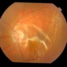
 Chorioretinal Folds Orbital Tumor Histo Path
Jun 20 2014 by Robert T. Wendel, MD
Chorioretinal folds orbital tumor.
Condition/keywords: chorioretinal fold
-
 Chorioretinal Folds Orbital Tumor
-
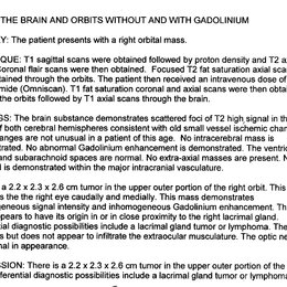 MRI Chorioretinal Folds
Jun 20 2014 by Robert T. Wendel, MD
Chorioretinal folds orbital tumor.
Condition/keywords: chorioretinal fold
-
---thumb.jpg/image-square;max$300,300.ImageHandler) Congenital Retinal Folds
Congenital Retinal Folds
Nov 6 2013 by Maurice F. Rabb
13 year old white male with congenital retinal folds.
Condition/keywords: congenital retinal folds
-
 Choroidal Folds
Nov 28 2014 by Thomas A. Ciulla, MD, MBA, FASRS
This 53 -year-old man was noted to have choroidal folds right greater than left. The visual acuity was normal at 20/15. The choroidal folds are visible on OCT, especially on the vertical cuts that image across the horizontal folds. Angiography revealed staining of the folds without CNVM, choroidal mass, or optic nerve edema.
Condition/keywords: bilateral chorioretinal folds, choroidal folds
-
---thumb.jpg/image-square;max$300,300.ImageHandler) Congenital Retinal Folds
Congenital Retinal Folds
Nov 6 2013 by Maurice F. Rabb
13 year old white male with congenital retinal folds.
Condition/keywords: congenital retinal folds
-
 Scleritis with Chorioretinal Folds
Feb 13 2015 by David Callanan, MD
46-year-old white male, scleritis with chorio-retinal folds.
Condition/keywords: chorioretinal fold, scleritis
-
---thumb.jpg/image-square;max$300,300.ImageHandler) Congenital Retinal Folds
Congenital Retinal Folds
Nov 6 2013 by Maurice F. Rabb
13 year old white male with congenital retinal folds.
Condition/keywords: congenital retinal folds
-
 Chorioretinal Folds Orbital Tumor
Jun 20 2014 by Robert T. Wendel, MD
Chorioretinal folds orbital tumor.
Condition/keywords: chorioretinal fold
-
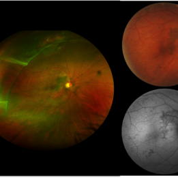
 Hypotony Maculopathy
Apr 1 2019 by Anfisa Ayalon, MD
Fundus autofluorescence image of 81-year-old male with right eye ocular hypotony due to leaking bleb. Note severe hypotony maculopathy, peripheral choroidal detachments, multiple chorioretinal folds.
Photographer: Anfisa Ayalon, MD., Meir Medical Center, Kfar Saba, Israel.
Imaging device: California, Optos 200 DTX
Condition/keywords: choroidal detachment, choroidal folds, fundus autofluorescence (FAF), hypotonous retinopathy, hypotony maculopathy
-
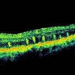 Retinal Folds Following Retinal Reattachment Surgery
Nov 22 2015 by Mallika Goyal, MD
Multiple retinal folds 4 weeks following vitreous surgery (perfluorodecalin assisted) for retinal detachment with giant retinal tear. OCT shows residual subretinal fluid and outer retinal folds (ORFs) seen as vertical hyperreflective lesions consisting of folded inner segment/outer segment of photoreceptors band and external limiting membrane band.
Photographer: Mallika Goyal, MD, Apollo Health City, Jubilee Hills, Hyderabad, India
Condition/keywords: retinal fold
-
 Choroidal Folds
Nov 28 2014 by Thomas A. Ciulla, MD, MBA, FASRS
This 53-year-old man was noted to have choroidal folds right greater than left. The visual acuity was normal at 20/15. The choroidal folds are visible on OCT, especially on the vertical cuts that image across the horizontal folds. Angiography revealed staining of the folds without CNVM, choroidal mass, or optic nerve edema.
Photographer: Charlotte Harris
Condition/keywords: bilateral chorioretinal folds, choroidal folds
-
---thumb.jpg/image-square;max$300,300.ImageHandler) Congenital Retinal Folds
Congenital Retinal Folds
Nov 6 2013 by Maurice F. Rabb
13 year old white male with congenital retinal folds.
Condition/keywords: congenital retinal folds
-
---thumb.jpg/image-square;max$300,300.ImageHandler) Congenital Retinal Folds
Congenital Retinal Folds
Nov 6 2013 by Maurice F. Rabb
13 year old white male with congenital retinal folds.
Condition/keywords: congenital retinal folds
-
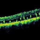 Retinal Folds Following Retinal Reattachment Surgery
Nov 22 2015 by Mallika Goyal, MD
Multiple retinal folds 4 weeks following vitreous surgery (perfluorodecalin assisted) for retinal detachment with giant retinal tear. OCT shows residual subretinal fluid and outer retinal folds (ORFs) seen as vertical hyperreflective lesions consisting of folded inner segment/outer segment of photoreceptors band and external limiting membrane band.
Photographer: Mallika Goyal, MD, Apollo Health City, Jubilee Hills, Hyderabad, India
Condition/keywords: retinal fold
 Loading…