 Initializing download.
-
 By Thomas A. Ciulla, MD, MBA, FASRS
Indiana University School of Medicine - Uploaded on Nov 28, 2014.
- Last modified by Caroline Bozell on Dec 1, 2014.
- Rating
- Appears in
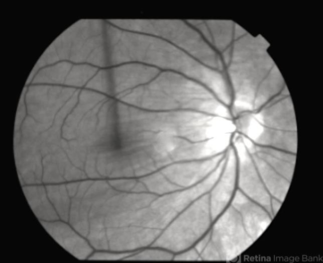
- Choroidal Folds
- Condition/keywords
- choroidal folds, bilateral chorioretinal folds
- Photographer
- Charlotte Harris
- Imaging device
- Fundus camera
- Description
- This 53-year-old man was noted to have choroidal folds right greater than left. The visual acuity was normal at 20/15. The choroidal folds are visible on OCT, especially on the vertical cuts that image across the horizontal folds. Angiography revealed staining of the folds without CNVM, choroidal mass, or optic nerve edema.


