Search results (100 results)
-
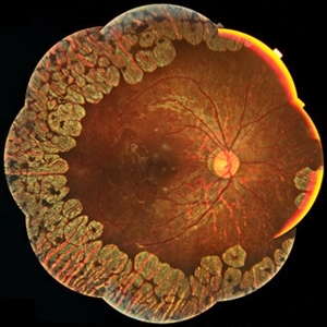
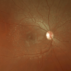
 Reticular Drusen, Doyne's Honeycomb Retinal Dystrophy, Malattia Leventinese, Familial Dominant Drusen
Feb 22 2018 by Nichole Lewis
Reticular Drusen, Doyne's Honeycomb Retinal Dystrophy, Malattia Leventinese, Familial Dominant Drusen
Photographer: Nichole Lewis
Condition/keywords: Doyne's Honeycomb, Familial Dominant Drusen, Malattia Leventinese, reticular drusen
-
 Gyrate Atrophy
Jan 6 2019 by Hashim Ali Khan, OD, FAAO
Montage of Multiple Fundus Photographs from the right eye of a 25-year-old woman with gyrate atrophy.
Photographer: Ahmed Abbass
Imaging device: Topcon TRC-NW8F
Condition/keywords: gyrate atrophy, hereditary retinal dystrophy, retinal dystrophy
-
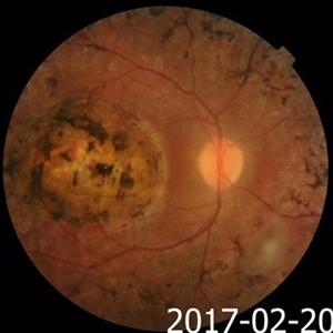
 Central Areolar Choroidal Dystrophy
Apr 14 2018 by Hamza Ahmed Shawky
Right fundus color photograph of a 35-year-old man with central areolar choroidal dystrophy, BCVA is 6/60
Photographer: Hamza Shawky, Alferdaws eye hospital, Retina unit
Imaging device: Heidelberg Spectralis
Condition/keywords: central areolar choroidal dystrophy (CACD), hereditary retinal dystrophy, macular dystrophy, retinal dystrophy
-
 Reticular Drusen, Doyne's Honeycomb Retinal Dystrophy, Malattia Leventinese, Familial Dominant Drusen
Feb 22 2018 by Nichole Lewis
Reticular Drusen, Doyne's Honeycomb Retinal Dystrophy, Malattia Leventinese, Familial Dominant Drusen
Photographer: Nichole Lewis
Condition/keywords: Doyne's Honeycomb, Familial Dominant Drusen, Malattia Leventinese, reticular drusen
-
 Stargardt's Disease LE
Jun 4 2014 by Neha Goel, MS DNB FRCS (Glasg)
Fundus photograph of the left eye of a 20-year-old male.
Photographer: Neha Goel
Imaging device: Zeiss Visucam
Condition/keywords: hereditary retinal dystrophy, Stargardt disease
-
---thumb.jpg/image-square;max$300,300.ImageHandler) Retinal Dystrophy
Retinal Dystrophy
Aug 9 2013 by From the Collections of Thomas M. Aaberg, MD and Thomas M. Aaberg Jr., MD
Pigmented dystrophy.
Condition/keywords: pigmentary retinal dystrophy, retinal dystrophy
-
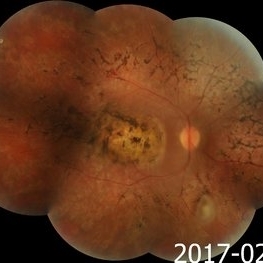
 Macular Coloboma and Pigmentary Retinopathy
Feb 25 2017 by Hamid Ahmadieh, MD
Color fundus photograph of the right eye of a 25-year-old woman with the history of low vision since childhood. Bilateral macular colobomata and pigmentary retinopathy similar to Leber's congenital amaurosis are present.
Photographer: Shabnam Poureh, Negah Eye Center, Tehran, Iran
Condition/keywords: bilateral pigmentary retinopathy, color fundus photograph, macular coloboma, pigmentary retinal dystrophy
-
 Retinitis Pigmentosa
Apr 30 2015 by Mitzy E Torres Soriano, MD
Fundus of patient with retinitis pigments, bone spicule-shaped pigment deposits are present with retinal atrophy, while the macula is preserved . Retinal vessels are attenuated.
Photographer: Mitzy E. Torres Soriano, MD; Centro medico Cagua-Estado Aragua. Venezuela
Imaging device: TRC-NW8
Condition/keywords: pigmentary retinal dystrophy, retinal dystrophy, retinitis pigmentosa, retinitis pigmentosa (RP) dystrophy
-
 Stargardt's Disease RE
Jun 4 2014 by Neha Goel, MS DNB FRCS (Glasg)
Fundus photograph of the right eye of a 20-year-old male.
Photographer: Neha Goel
Imaging device: Zeiss Visucam
Condition/keywords: hereditary retinal dystrophy, Stargardt disease
-
---thumb.jpg/image-square;max$300,300.ImageHandler) Retinal Dystrophy
Retinal Dystrophy
Aug 9 2013 by From the Collections of Thomas M. Aaberg, MD and Thomas M. Aaberg Jr., MD
Large PED.
Condition/keywords: pigment epithelial detachment (PED), retinal dystrophy
-
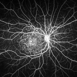
 Retinal Dystrophy of 24-Year-Old Male/ AF OD
Nov 25 2015 by Zach Dupureur
Fluorescein angiography of a 24-year-old male. Juvenile retinoschisis on OCT. FA shows outer retinal staining. Could be associated with Goldman Farve Syndrome.
Photographer: Zach Dupureur OCT-C
Imaging device: Heidelberg Spectralis
Condition/keywords: Goldmann-Favre Syndrome, juvenile retinoschisis, retinal dystrophy
-
 Central Areolar Choroidal Dystrophy
Apr 14 2018 by Hamza Ahmed Shawky
Left fundus color photograph of a 35-year-old man with central areolar choroidal dystrophy, BCVA is 6/60.
Photographer: Hamza Shawky, Alferdaws eye hospital, Retina unit
Imaging device: Heidelberg Spectralis
Condition/keywords: central areolar choroidal dystrophy (CACD), hereditary retinal dystrophy, macular dystrophy, retinal dystrophy
-
 Pigmentary Retinal Dystrophy
Feb 9 2015 by Matt Poe, COA
This was a lady that presented with bilateral pigmentary retinal dystrophy.
Photographer: Matt Poe, COA. Northwest Arkansas Retina Associates, Springdale, AR.
Condition/keywords: hereditary retinal dystrophy, pigmentary retinal dystrophy
-
 Stargardt's Disease
Aug 20 2015 by Cory Mangham
Fundus photograph of 27-year-old female with Stargardt's disease.
Photographer: Cory Mangham CRA
Imaging device: Optos 200Tx
Condition/keywords: retinal dystrophy, Stargardt disease
-
 Stargard's Disease
Aug 20 2015 by Cory Mangham
Fluorescein angiogram of 27-year-old female with Stargardt's disease.
Photographer: Cory Mangham CRA
Imaging device: Optos 200Tx
Condition/keywords: retinal dystrophy, Stargardt disease
-
---thumb.jpg/image-square;max$300,300.ImageHandler) Retinal Dystrophy
Retinal Dystrophy
Aug 9 2013 by From the Collections of Thomas M. Aaberg, MD and Thomas M. Aaberg Jr., MD
FA of central atrophy with flecks.
Condition/keywords: central choroidal atrophy, total, flecks, retinal dystrophy
-
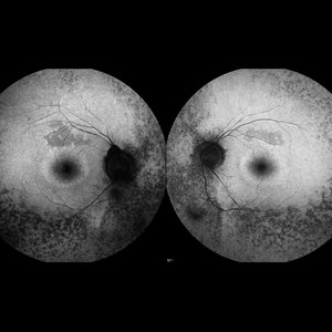 Retinitis Pigmentosa
May 27 2016 by Olivia Rainey
Bilateral fundus autofluorescence images of retinitis pigmentosa.
Photographer: Olivia Rainey
Imaging device: Heidelberg Spectralis
Condition/keywords: 50 degrees, bilateral, fundus autofluorescence (FAF), hereditary retinal dystrophy, retinitis pigmentosa
-
 Pigmentary Retinal Dystrophy
Mar 29 2019 by Jessica Norkus
Optos ultra wide field image of 41-year-old male patient with pigmentary retinal dystrophy. Atypical findings due to unilateral presentation. Patient has been experiencing symptoms for 15 years, notes significant nyctalopia.
Photographer: Jessica Norkus
Imaging device: Optos Ultra Wide Field Camera
Condition/keywords: abnormal fundus, bone spicule, color fundus photograph, color photo, fundus photograph, Optos, peripheral bone spicules, pigment changes, ultra-wide field imaging, unilateral blindness
-
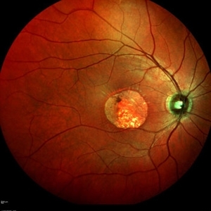
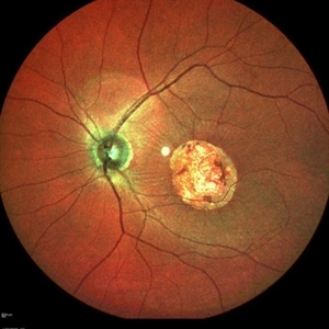
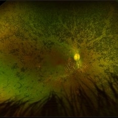
 Doyne Honeycomb Retinal Dystrophy
Sep 29 2020 by Navneet Mehrotra, DNB
Left eye fundus photograph of a 36-year-old female with decreased vision both eyes for six months. Father also had a similar retinal disorder.
Photographer: Dr Navneet Mehrotra
Imaging device: TRC- NW8F
Condition/keywords: Doyne's Honeycomb, drusen, Malattia Leventinese
-
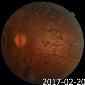
 Macular Coloboma and Pigmentary Retinopathy
Feb 25 2017 by Hamid Ahmadieh, MD
Merged color fundus photograph of the right eye of a 25-year-old woman with the history of low vision since childhood. Bilateral macular colobomata and pigmentary retinopathy similar to Leber's congenital amaurosis are present.
Photographer: Shabnam Poureh, Negah Eye Center, Tehran, Iran
Condition/keywords: bilateral pigmentary retinopathy, color fundus photograph, macular coloboma, pigmentary retinal dystrophy
-
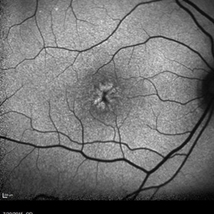
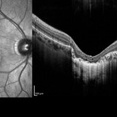
 Central Areolar Choroidal Dystrophy
Apr 14 2018 by Hamza Ahmed Shawky
Left fundus OCT of a 35-year-old man with central areolar choroidal dystrophy, BCVA is 6/60
Photographer: Hamza Shawky, Alferdaws eye hospital, Retina unit
Imaging device: Heidelberg Spectralis
Condition/keywords: central areolar choroidal dystrophy (CACD), hereditary retinal dystrophy, macular dystrophy, retinal dystrophy
-
---thumb.jpg/image-square;max$300,300.ImageHandler) Stargardt 's Disease
Stargardt 's Disease
Jun 27 2013 by Raj K. Maturi, MD
FAF of a 30-year-old male with hereditary retinal dystrophy.
Photographer: Charlotte Harris COA Midwest Eye Institute Indianapolis, IN
Imaging device: Heidelberg Spectralis
Condition/keywords: Stargardt disease
-
---thumb.jpg/image-square;max$300,300.ImageHandler) Retinal Dystrophy
Retinal Dystrophy
Aug 9 2013 by From the Collections of Thomas M. Aaberg, MD and Thomas M. Aaberg Jr., MD
Pigmented dystrophy.
Condition/keywords: pigmentary retinal dystrophy, retinal dystrophy
-
 Macular Coloboma and Pigmentary Retinopathy
Feb 25 2017 by Hamid Ahmadieh, MD
Color fundus photograph of the right eye of a 25-year-old woman with the history of low vision since childhood. Bilateral macular colobomata and pigmentary retinopathy similar to Leber's congenital amaurosis are present.
Photographer: Shabnam Poureh, Negah Eye Center, Tehran, Iran
Condition/keywords: color fundus photograph, pigmentary retinal dystrophy
-
 Central Areolar Choroidal Dystrophy
Apr 14 2018 by Hamza Ahmed Shawky
Right fundus OCT of a 35-year-old man with central areolar choroidal dystrophy, BCVA is 6/60
Photographer: Hamza Shawky, Alferdaws eye hospital, Retina unit
Imaging device: Heidelberg Spectralis
Condition/keywords: central areolar choroidal dystrophy (CACD), hereditary retinal dystrophy, macular dystrophy, retinal dystrophy
 Loading…