Search results (35 results)
-
 Toxoplasmosis Slide 2
Oct 22 2012 by Ronald C. Gentile, MD
One month following treatment with Bactrim, Clindamycin, and oral prednisone the focal area chorioretinitis has coalesced with a decrease in overlying vitreous inflammation. Kyrieleis plaques can be seen along the inferior retinal arteriole.
Photographer: The New York Eye & Ear Infirmary Department of Medical Imaging
Condition/keywords: posterior uveitis, toxoplasmosis
-
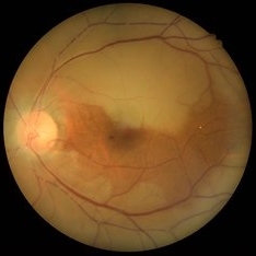
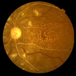 Primary Hyperoxaluria and Oxalosis
Oct 10 2015 by Hamid Ahmadieh, MD
Color fundus photograph of the left eye of a 55-year-old woman with primary hyperoxaluria and oxalosis leading to intraretinal and subretinal deposition of calcium oxalate crystals . In addition, deposition of these crystals in the retinal vessels has led to the occlusion of retinal arterioles and venules leading to multiple cotton wools and dot and blot retinal hemorrhages.
Photographer: Shabnam Pooreh, Negah Eye Center, Tehran, Iran
Condition/keywords: color fundus photograph, oxalosis, primary hyperoxaluria
-
---thumb.jpg/image-square;max$300,300.ImageHandler) Brown/Mendis BJO 57:344, 1973
Brown/Mendis BJO 57:344, 1973
Feb 14 2013 by From the Collections of Thomas M. Aaberg, MD and Thomas M. Aaberg Jr., MD
reprints of figures 1 and 2 from the publication Brown and Mendis. Retinal arteritis complicating herpes zoster ophthalmicus. Br J Ophthalmol 1973;57:344-6. The left panel is a "fundus painting showing extensive exudate in areas of supply of narrowed and sheathed upper nasal and upper temporal retinal arterioles." The right panel is a fluorescein angiograph of the fundus, "demonstrating leakage of dye in area of exudation."
Condition/keywords: Herpes zoster, retinal arteriolar occlusion, retinal necrosis
-
 Lignocaine Retinal Toxicity
Aug 18 2015 by Mallika Goyal, MD
Right eye fundus of a 70-year-old male 3 weeks after inadvertent globe penetration with peribulbar anaesthesia needle and intraocular injection of lignocaine. There is a thick taut epimacular membrane with severely increased central retinal thickness. Fluorescein angiography revealed an occluded retinal arteriole at the macula indicating macular ischaemia underlying the membrane.
Photographer: Mallika Goyal, MD, Apollo Health City, Jubilee Hills, Hyderabad
Condition/keywords: lignocaine retinal toxicity
-
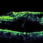 Lignocaine Retinal Toxicity
Aug 21 2015 by Mallika Goyal, MD
Right eye OCT of a 70-year-old male 3 weeks after inadvertent globe penetration with peribulbar anaesthesia needle and intraocular injection of lignocaine. There is a thick taut epimacular membrane with severely increased central retinal thickness. Fluorescein angiography revealed an occluded retinal arteriole at the macula indicating macular ischaemia underlying the membrane.
Photographer: Mallika Goyal, MD, Apollo Health City, Jubilee Hills, Hyderabad, India
Condition/keywords: lignocaine retinal toxicity
-
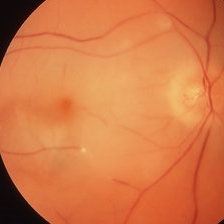
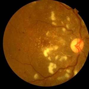 Primary Hyperoxaluria and Oxalosis
Oct 10 2015 by Hamid Ahmadieh, MD
Color fundus photograph of the right eye of a 55-year-old woman with primary hyperoxaluria and oxalosis leading to intraretinal and subretinal deposition of calcium oxalate crystals . In addition, deposition of these crystals in the retinal vessels has led to the occlusion of retinal arterioles and venules leading to multiple cotton wools and dot and blot retinal hemorrhages.
Photographer: shabnam Pooreh, Negah Eye Center, Tehran, Iran
Condition/keywords: color fundus photograph, oxalosis, primary hyperoxaluria
-
 Lignocaine Retinal Toxicity
Aug 18 2015 by Mallika Goyal, MD
Right eye fundus of a 70-year-old male 3 weeks after inadvertent globe penetration with peribulbar anaesthesia needle and intraocular injection of lignocaine. There is a thick taut epimacular membrane with severely increased central retinal thickness. Fluorescein angiography revealed an occluded retinal arteriole at the macula indicating macular ischaemia underlying the membrane.
Photographer: Mallika Goyal, MD, Apollo Health City, Jubilee Hills, Hyderabad
Condition/keywords: lignocaine retinal toxicity
-
 Lignocaine Retinal Toxicity
Aug 18 2015 by Mallika Goyal, MD
Right eye fluorescein angiogram of a 70-year-old male 3 weeks after inadvertent globe penetration with peribulbar anaesthesia needle and intraocular injection of lignocaine. There is an occluded retinal arteriole indicating macular ischaemia underlying the clinically obvious epimacular membrane.
Photographer: Mallika Goyal, MD, Apollo Health City, Jubilee Hills, Hyderabad
Condition/keywords: lignocaine retinal toxicity
-
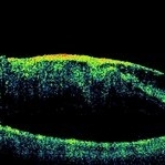
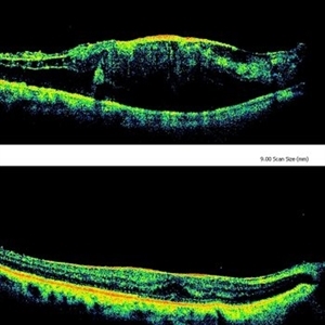 Lignocaine Retinal Toxicity
Aug 21 2015 by Mallika Goyal, MD
Right eye OCT of a 70-year-old male 3 weeks after inadvertent globe penetration with peribulbar anaesthesia needle and intraocular injection of lignocaine showing a taut epimacular membrane with macular elevation compared to a relatively normal foveal contour immediately after surgery suggesting progressive traction secondary to lignaocaine toxicity. Fluorescein angiography revealed an occluded retinal arteriole at the macula indicating macular ischaemia underlying the membrane.
Photographer: Mallika Goyal, MD, Apollo Health City, Jubilee Hills, Hyderabad, India
Condition/keywords: lignocaine retinal toxicity
-
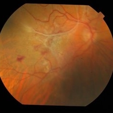
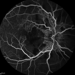 Primary Hyperoxaluria and Oxalosis
Oct 10 2015 by Hamid Ahmadieh, MD
Late venous phase FA image of the right of a 55-year-old woman with primary hyperoxaluria and oxalosis . Notice macular infarction and areas of capillary non -perfusion in retinal mid periphery due to the occlusion of retinal arterioles.
Photographer: Shabnam Pooreh, Negah Eye Center, Tehran, Iran
Condition/keywords: oxalosis, primary hyperoxaluria
-
 Lignocaine Retinal Toxicity
Aug 21 2015 by Mallika Goyal, MD
Right eye OCT of a 70-year-old male 3 weeks after inadvertent globe penetration with peribulbar anaesthesia needle and intraocular injection of lignocaine shows a thick taut epimacular membrane with severely increased central retinal thickness. Fluorescein angiography revealed an occluded retinal arteriole at the macula indicating macular ischaemia underlying the membrane.
Photographer: Mallika Goyal, MD, Apollo Health City, Jubilee Hills, Hyderabad, India
Condition/keywords: lignocaine retinal toxicity
-
 Lignocaine Retinal Toxicity
Aug 18 2015 by Mallika Goyal, MD
Right eye fluorescein angiogram of a 70-year-old male 3 weeks after inadvertent globe penetration with peribulbar anaesthesia needle and intraocular injection of lignocaine. There is an occluded retinal arteriole indicating macular ischaemia underlying the clinically obvious epimacular membrane.
Photographer: Mallika Goyal, MD, Apollo Health City, Jubilee Hills, Hyderabad
Condition/keywords: lignocaine retinal toxicity
-
 Slide 9-20
Feb 26 2019 by Lancaster Course in Ophthalmology
Cholesterol emboli to retina and choroid. There is a large microinfarction of the nerve fiber layer (arrows). A cholesterol embolus is lodged in a retinal arteriole (upper right) proximal to the microinfarction. Cholesterol emboli were also found in the choroid, and in one (lower right) erythrocytes (arrow) could be seen in the periphery as they were presumably going around the embolus.
Condition/keywords: choroid, emboli, embolus, erythrocytes
-
 Slide 9-18
Feb 26 2019 by Lancaster Course in Ophthalmology
Malignant hypertension with retinal arterioles that are thickened and have fibrinoid necrosis (arrows). Retinal exudates (asterisk) and papilledema are also present. Papilledema is evidenced by fullness of the optic nerve head and peripapillary crowding of the retina (lower right).
Condition/keywords: fibrinoid, malignant hypertension, papilledema, retinal arteriole, retinal exudates
-
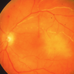
 Central Retinal Artery Occlusion with Cilioretinal Sparing
Oct 28 2020 by Fang Helen Mi
Fundus photograph of an 61-year-old Chinese male showing central retinal artery occlusion with cilioretinal sparing. Photo shows diffuse ischemic retinal whitening and box-carring of the retinal arterioles.
Condition/keywords: central retinal artery occlusion (CRAO), cilioretinal sparing
-
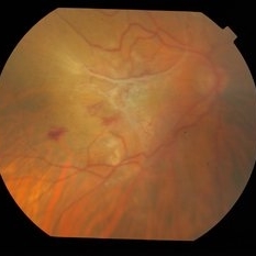
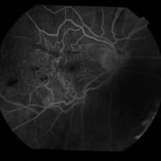
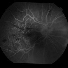
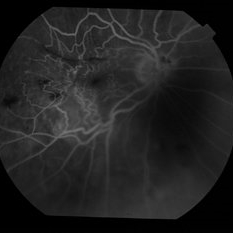
 Idiopathic retinal vasculitis, aneurysms and neuroretinitis
Apr 24 2022 by Aniruddha K Agarwal, MD
Ultra-wide field fundus fluorescein angiography (FFA) of the left eye from an asymptomatic, healthy 33-year-old woman who was referred to the retina clinic from a refractive surgery unit due to the presence of vascular anomalies and hard exudates in both eyes. FFA revealed the characteristic sacular aneurysms at the bifurcation of retinal arterioles in the posterior pole, together with microvascular anomalies and capillary closure peripherally.
Photographer: Julio J GONZALEZ-LOPEZ, MD, PhD, FEBO and Teresa GONZALEZ-LOMAS, RN
Imaging device: Optos California
Condition/keywords: IRVAN Syndrome, IUSG, neuroretinitis, retinal vasculitis, uveitis
-
 Embolic Central Retinal Artery Occlusion
Mar 26 2019 by Gary R. Cook, MD, FACS
58-year-old WM with embolic CRAO demonstrating a a cherry-red spot in macula, retinal whitening around the fovea, and the embolus in a inferotemporal branch retinal arteriole; VA= HM 6''
Imaging device: Topcon VT-50
Condition/keywords: central retinal artery occlusion (CRAO), cherry red spot, embolus, retinal whitening
-
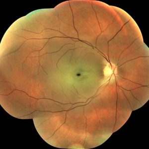
 Slide 4-6
Feb 20 2019 by Lancaster Course in Ophthalmology
Optic atrophy. Note the marked narrowing of the retinal arterioles.
Condition/keywords: optic atrophy, retinal arteriole
-
 Central Retinal Artery Occlusion
Mar 2 2021 by Renata Garcia Franco, Md
Retinal edema, cherry spot, retinal arteriolar attenuation and segmentation of blood in retinal arterioles.
Photographer: Guillermina Hernandez
Imaging device: Zeiss
Condition/keywords: central artery
-
 Central Retinal Artery Occlusion
Jan 22 2021 by Renata Garcia Franco, Md
65-year-old male, history of uncontrolled systemic arterial hypertension. Segmentation of blood in retinal arterioles, retinal whitening and cherry red spot.
Photographer: Fatima Hernandez, Instituto de la Retina del Bajio SC
Imaging device: Zeiss
Condition/keywords: central retinal artery occlusion (CRAO)
-
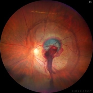 Ruptured Retinal Artery Macroaneurysm
Jun 18 2024 by KANWALJEET HARJOT MADAN, M.S. (Ophthalmology), FAICO (Vitreous - Retina)
This is a fundus photo depicting ruptured Retinal Artery Macroaneurysm (RAM) in the left eye of a 63 years old female. RAM is an acquired saccular or fusiform dilatation of the retinal arterioles that usually occur within the first three orders of bifurcation. The Superotemporal artery is the most common location. RAM may be asymptomatic or cause a number of complications such as macular edema, serous macular detachment, and hemorrhages.
Photographer: Dr Kanwaljeet Harjot Madan
Condition/keywords: Haemorrhage, macroaneurysm, retinal arteriole
-
 Bilateral Calcific Retina Arteriolar Occlusions in a Patient with Metastatic Ovarian Carcinoma
Dec 10 2020 by McGill University Health Centre
47-year-old female with cough and fever. Imaging showed a right pulmonary infiltrate. Transbronchial needle biopsy revealed lymphangitic spread of papillary adenocarcinoma with psammoma bodies. MRI of thyroid, CT of abdomen and pelvis were negative. gynecologic evaluation negative at that time . The patient had bilateral floaters, VA: 20/40 OD and 20/20 OS. Fundus examination showed retinal arteriolar sheathing and a flat choroidal lesion OS and vitritis OD. Fluorescein angiogram showed staining of left superior temporal retinal arterioles and bilateral midperipheral patchy hyperfluorescence at RPE The patient vision in the OD deteriorated to 20/400, and in the OS 20/50. Four months later a new choroidal lesion was diagnosed OS. An abdominal mass consistent with a cystadenoma of the ovary was diagnosed. After a year patient developed systemic metastasis. Autopsy: Metastatic adenocarcinoma to the lung, both adrenals, para-aortic lymph nodes, left hip, right breast, occipital skin, serosal surface of liver, pituitary. In almost all metastatic lesions psammoma bodies were found. Presumptive diagnosis is a primary tumor of the ovary.
Condition/keywords: bilateral, calcification, metastatic adenocarcinoma, retinal arteriolar occlusion
-
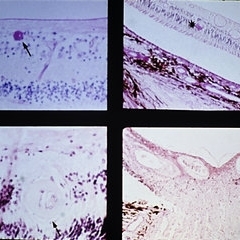
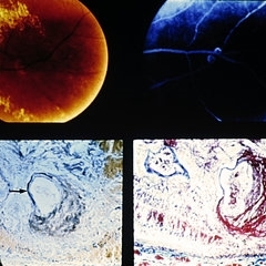 Slide 9-19
Feb 26 2019 by Lancaster Course in Ophthalmology
Retinal arterial macroaneurysm. A ring of retinal exudate partially surrounds the macroaneurysm (upper left), which is more clearly delineated by fluorescein (upper right). The retinal arteriole is greatly dilated, and the stain for elastic tissue shows a localized area of disruption and loss of the internal elastic membrane (arrow). The surrounding retina is thickened by edema and some hemorrhage. The ectatic area of the vessel wall is greatly thickened by the accumulation of a laminated fibrinous material. (Courtesy of Alan Friedman, M.D.)
Condition/keywords: retinal arterial macroaneurysm, retinal exudates
-
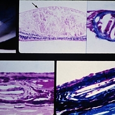
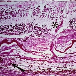 Slide 9-84
Feb 26 2019 by Lancaster Course in Ophthalmology
Senile macular degeneration with disciform scar. A retinal arteriole (asterisk) extends into the subretinal component of the scar, through a break in the thickened and detached inner layer of Bruch's membrane, and then into the vascularized intra-Bruch's-membrane component of the scar. Study of serial sections disclosed this retinal vessel to anastomose with the choroidal vessel (arrow) which extends through a branch in Bruch's membrane.
Condition/keywords: Bruch's membrane, disciform scar, macular degeneration, retinal arteriole
 Loading…