Search results (66 results)
-
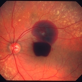
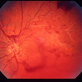
 "Boat-Shaped" Preretinal Hemorrhage
Feb 21 2019 by Mitzy E Torres Soriano, MD
Color fundus photograph showing preretinal (subhyaloid) hemorrhage in a diabetic patient with proliferative diabetic retinopathy.
Photographer: Andrea Vitale, MD
Condition/keywords: preretinal hemorrhage, proliferative diabetic retinopathy (PDR), subhyaloid hemorrhage
-
 PDR with Active NVD
Oct 8 2012 by Jeffrey G. Gross, MD, FASRS
PDR with active NVD and preretinal hemorrhage, mild VH and partial PRP.
Condition/keywords: neovascularization of the disc (NVD), preretinal hemorrhage, scatter laser photocoagulation, vitreous hemorrhage
-
 Ruptured retinal arterial macroaneurysm
Jan 11 2013 by Alex P. Hunyor, MD
Retinal arterial macroaneurysm with subretinal and preretinal hemorrhage
Condition/keywords: retinal arterial macroaneurysm
-
 Preretinal Hemorrhage - OCT
Sep 20 2012 by Allen Chiang, MD, FASRS
34-year old woman with preretinal hemorrhage in the macula, with dehemoglobinization occuring within the central portion of the hemorrhage while undergoing observation.
Imaging device: Zeiss Cirrus
Condition/keywords: preretinal hemorrhage
-
 Preretinal Hemorrhage
Sep 20 2012 by Allen Chiang, MD, FASRS
34-year old woman with preretinal hemorrhage in the macula, with dehemoglobinization occuring within the central portion of the hemorrhage while undergoing observation.
Imaging device: Zeiss Cirrus
Condition/keywords: preretinal hemorrhage
-
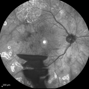
 Proliferative Diabetic Retinopathy
Sep 15 2012 by Hamid Ahmadieh, MD
Fundus autofluorescence image of a 30-year-old woman with the history of scatter laser photocoagulation and a preretinal hemorrhage due to active PDR .
Photographer: Hamid Ahmadieh, MD, Ophthalmic Research Center, Labbafinejad Medical Center, Shahid Beheshti University of Medical Sciences
Imaging device: Heidelberg HRA
Condition/keywords: fundus autofluorescence (FAF), preretinal hemorrhage
-
 PDR with Active NVD
Oct 8 2012 by Jeffrey G. Gross, MD, FASRS
PDR with active NVD and preretinal hemorrhage.
Condition/keywords: neovascularization of the disc (NVD), preretinal hemorrhage
-
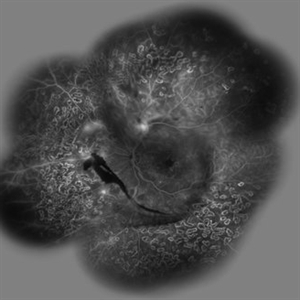 Proliferative Diabetic Retinopathy (PDR)
Sep 11 2012 by Hamid Ahmadieh, MD
Wide- field FA image of a 55-year-old woman with active PDR and the history of scatter laser photocoagulation.
Photographer: Hamid Ahmadieh, MD, Ophthalmic Research Center, Labbafinejad Medical Center, Shahid Beheshti University of Medical Sciences
Imaging device: Heidelberg HRA
Condition/keywords: preretinal hemorrhage, retinal neovascularization, scatter laser photocoagulation
-
 Scleral Indentation
Nov 9 2012 by Norman Byer
This is the same lesion with scleral indentation. You can see the small discrete preretinal hemorrhage and the sharply circumscribed area of elevated retina with subretinal fluid beneath it. No retinal break is visible, but the posterior vitreous is detached and exerting traction at this site. The area was surrounded with argon laser treatment the same day as the initial examination.
Condition/keywords: posterior vitreous detachment, preretinal hemorrhage, scleral indentation, subretinal fluid, vitreous traction
-
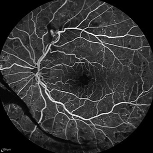 Proliferative Diabetic Retinopathy (PDR)
Sep 11 2012 by Hamid Ahmadieh, MD
FA image of a 55-year-old woman with active PDR.
Photographer: Hamid Ahmadieh, MD, Ophthalmic Research Center, Labbafinejad Medical Center, Shahid Beheshti University of Medical Sciences
Imaging device: Heidelberg HRA
Condition/keywords: preretinal hemorrhage, retinal neovascularization
-
---thumb.jpg/image-square;max$300,300.ImageHandler) Retinal Neovascularization
Retinal Neovascularization
Feb 13 2013 by From the Collections of Thomas M. Aaberg, MD and Thomas M. Aaberg Jr., MD
Lipid deposit, NVD.
Condition/keywords: lipid, neovascularization of the disc (NVD), preretinal hemorrhage, retinal neovascularization
-
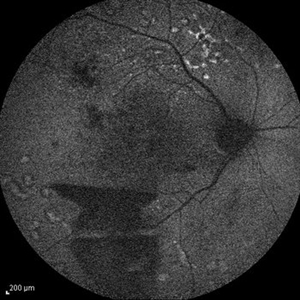
 Proliferative Diabetic Retinopathy
Sep 15 2012 by Hamid Ahmadieh, MD
Infrared image of a 30-year-old woman with the history of scatter laser photocoagulation and a preretinal hemorrhage due to active PDR .
Photographer: Hamid Ahmadieh, MD, Ophthalmic Research Center, Labbafinejad Medical Center, Shahid Beheshti University of Medical Sciences
Imaging device: Heidelberg HRA
Condition/keywords: infrared image, preretinal hemorrhage
-
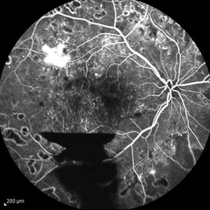 Proliferative Diabetic Retinopathy
Sep 15 2012 by Hamid Ahmadieh, MD
FA image of a 30-year-old woman with the history of scatter laser photocoagulation, NVE and a preretinal hemorrhage due to active PDR .
Photographer: Hamid Ahmadieh, MD, Ophthalmic Research Center, Labbafinejad Medical Center, Shahid Beheshti University of Medical Sciences
Imaging device: Heidelberg HRA
Condition/keywords: preretinal hemorrhage
-
 Preretinal Hemorrhage due to Proliferative Diabetic Retinopathy
Oct 17 2012 by Sharon Fekrat, MD FACS FASRS
Fluorescein angiography of right eye with preretinal hemorrhage from neovascularization elsewhere associated with proliferative diabetic retinopathy. Note associated fibrosis.
Photographer: John Reaves, Ophthalmic Photographer, Durham VA Medical Center, Durham, NC
Condition/keywords: preretinal hemorrhage
-
---thumb.jpg/image-square;max$300,300.ImageHandler) Preretinal Hemorrhage in Proliferative Diabetic Retinopathy
Preretinal Hemorrhage in Proliferative Diabetic Retinopathy
Oct 17 2012 by Sharon Fekrat, MD FACS FASRS
Fluorescein angiography of right eye with preretinal hemorrhage from neovascularization elsewhere associated with proliferative diabetic retinopathy.
Photographer: John Reaves, Ophthalmic Photographer, Durham VA Medical Center, Durham, NC
Imaging device: Fluorescein angiography
Condition/keywords: preretinal hemorrhage, subhyaloid hemorrhage
-
 Regressed Proliferative Diabetic Retinopathy with Preretinal Hemorrhage
Oct 17 2012 by Sharon Fekrat, MD FACS FASRS
Fundus photograph of left eye of patient with regressed proliferative diabetic retinopathy following panretinal laser photocoagulation. Note fibrovascular proliferation along arcades and associated preretinal hemorrhage.
Photographer: John Reaves, Ophthalmic Photographer, Durham VA Medical Center, Durham, NC
Condition/keywords: preretinal hemorrhage, regressed
-
 Laser Photocoagulation
Nov 9 2012 by Norman Byer
This shows the same lesion four days after laser photo coagulation. The new hemorrhages seen in this photograph did not occur during the photocoagulation but developed within the next four days.
Condition/keywords: argon photocoagulation, laser photocoagulation, preretinal hemorrhage
-
---thumb.jpg/image-square;max$300,300.ImageHandler) Retinal Neovascularization
Retinal Neovascularization
Feb 13 2013 by From the Collections of Thomas M. Aaberg, MD and Thomas M. Aaberg Jr., MD
Preretinal hemorrhage neovascularization, dot-bbt hemorrhage.
Condition/keywords: preretinal hemorrhage, retinal neovascularization
-
---thumb.jpg/image-square;max$300,300.ImageHandler) Retinal Neovascularization
Retinal Neovascularization
Feb 13 2013 by From the Collections of Thomas M. Aaberg, MD and Thomas M. Aaberg Jr., MD
Lipid deposit, NVD.
Condition/keywords: lipid, neovascularization of the disc (NVD), preretinal hemorrhage, retinal neovascularization
-
 Preretinal Hemorrhage
May 6 2017 by Mitzy E Torres Soriano, MD
Fundus photograph of a 36-year-old-woman with a preretinal subhyaloid hemorrhage (valsalva retinopathy).
Photographer: Mitzy Torres Soriano
Condition/keywords: macular hemorrhage, premacular hemorrhage, preretinal hemorrhage, subhyaloid hemorrhage, valsalva retinopathy
-
 Dengue Fever
Oct 25 2012 by Mallika Goyal, MD
Fundus photograph of the left eye of a 32-year-old gentleman with dengue fever and thrombocytopenia. Photograph shows extensive retinal and pre-retinal haemorrhages, roth spots but no dengue retinitis. Same patient as in images 1-5.
Condition/keywords: Dengue Fever, preretinal hemorrhage, rosacea conjunctivitis
-
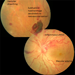 Inflammatory BRVO, Neovascularization and Macular Edema in a Patient with Vasculitis
Apr 4 2016 by AASHRAYA KARPE, MBBS, MS, FMRF
Composite fundus photograph of the left eye of a 20-year-old male with history of blurring of vision. Left eye revealed disc edema with a dilated tortuous superotemporal veins and hemorrhages in that quadrant. Mulitple preretinal hemorrhages were seen nasally along with vascular sheathing.
Imaging device: Zeiss FF450plus Fundus Camera
Condition/keywords: branch retinal vein occlusion (BRVO), macular edema, neovascularization (NV), subhyaloid hemorrhage, vascular sheathing of retina
-
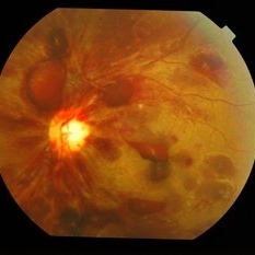
 BRVO Complications
Mar 29 2013 by Henry J. Kaplan, MD
Old superotemporal BRVO as a sclerotic vessel with NVD and NVE and vitreous hemorrhage and a preretinal hemorrhage.
Condition/keywords: branch retinal vein occlusion (BRVO), neovascularization (NV), neovascularization of the disc (NVD), vitreous hemorrhage
-
 Post-Op Day 10 Barrier Laser
Feb 13 2013 by From the Collections of Thomas M. Aaberg, MD and Thomas M. Aaberg Jr., MD
Barrier laser, preretinal hemorrhage.
Condition/keywords: barrier laser, post-op, preretinal hemorrhage
-
 Without Scleral Indentation
Nov 9 2012 by Norman Byer
This 54-year-old woman presented with a history of sudden light flashes and black floaters for five days. Without scleral indentation, her right eye showed this lesion at 10:30 just 4 disc diameters from the macular. This slide is the first of a series of six photographs showing the lesion in different stages.
Condition/keywords: black floaters, preretinal hemorrhage, without scleral indentation
 Loading…