Search results (59 results)
-
---thumb.jpg/image-square;max$300,300.ImageHandler) vitreous snowballs, inferior snowbanking, vascular sheathing
vitreous snowballs, inferior snowbanking, vascular sheathing
Feb 14 2013 by From the Collections of Thomas M. Aaberg, MD and Thomas M. Aaberg Jr., MD
schematic drawing of vitreous snowballs, inferior snowbanking, vascular sheathing
Condition/keywords: pars planitis, snowbank
-
 Pars Planitis - Peripheral Uveitis
Nov 9 2012 by Norman Byer
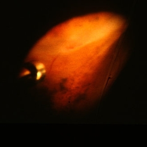
This 25-year-old man had pars planitis, peripheral uveitis bilaterally. In this eye it produced a small tractional oval tear of the retina and an inferior retinal detachment. The typical creamy yellow exudates of pars planitis can be seen in the lower right very close to the ora serrata.
Condition/keywords: creamy yellow exudates, inferior retinal detachment, pars planitis, peripheral uveitis, tractional retinal tear
-
---thumb.jpg/image-square;max$300,300.ImageHandler) vitreous snowballs, peripheral retinal neovascularization, inferior snowbanking, vascular sheathing, and peripheral exudative retinal detachment
vitreous snowballs, peripheral retinal neovascularization, inferior snowbanking, vascular sheathing, and peripheral exudative retinal detachment
Feb 14 2013 by From the Collections of Thomas M. Aaberg, MD and Thomas M. Aaberg Jr., MD
schematic drawing of vitreous snowballs, peripheral retinal neovascularization, inferior snowbanking, vascular sheathing, and peripheral exudative retinal detachment
Condition/keywords: exudative retinal detachment, pars planitis, peripheral retinal neovascularization, snowbank
-
---thumb.jpg/image-square;max$300,300.ImageHandler) inferior peripheral snowbanking
inferior peripheral snowbanking
Feb 14 2013 by From the Collections of Thomas M. Aaberg, MD and Thomas M. Aaberg Jr., MD
intraoperative indirect ophthalmoscopic photograph showing inferior peripheral snowbanking
Condition/keywords: pars planitis, snowbank
-
---thumb.jpg/image-square;max$300,300.ImageHandler) vitreous snowballs, peripheral retinal neovascularization, inferior snowbanking, and vascular sheathing
vitreous snowballs, peripheral retinal neovascularization, inferior snowbanking, and vascular sheathing
Feb 14 2013 by From the Collections of Thomas M. Aaberg, MD and Thomas M. Aaberg Jr., MD
schematic drawing of vitreous snowballs, peripheral retinal neovascularization, inferior snowbanking, and vascular sheathing
Condition/keywords: pars planitis, peripheral retinal neovascularization, snowbank
-
 peripheral pre-retinal fibrosis and neovascularization with vitreous cutter
Feb 14 2013 by From the Collections of Thomas M. Aaberg, MD and Thomas M. Aaberg Jr., MD
intraoperative photo of peripheral pre-retinal fibrosis and neovascularization with vitreous cutter
Condition/keywords: pars planitis, peripheral retinal neovascularization
-
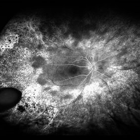
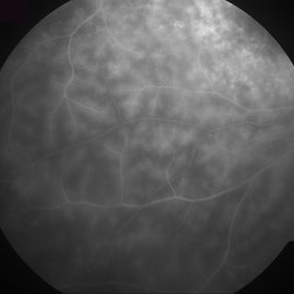
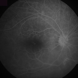
 pars planitis; macular cysts and serous detachment
Feb 14 2013 by From the Collections of Thomas M. Aaberg, MD and Thomas M. Aaberg Jr., MD
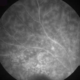
color and FA showing cystoid macular edema
Condition/keywords: cystoid macular edema (CME), pars planitis
-
---thumb.jpg/image-square;max$300,300.ImageHandler) peripheral pre-retinal fibrosis and neovascularization with vitreous cutter
peripheral pre-retinal fibrosis and neovascularization with vitreous cutter
Feb 14 2013 by From the Collections of Thomas M. Aaberg, MD and Thomas M. Aaberg Jr., MD
intraoperative photo of peripheral pre-retinal fibrosis and neovascularization with vitreous cutter
Condition/keywords: pars planitis, peripheral retinal neovascularization
-
 Epiretinal Membrane
Mar 27 2014 by Jason S. Calhoun
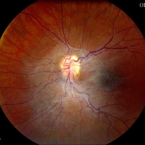
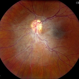
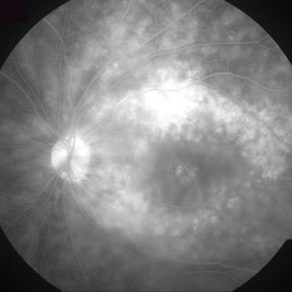
Patient with a history of pars planitis comes in with decreased vision in the left eye. VA is 20/30 in the left eye. Fundus photos show a rather large epiretinal membrane covering the optic nerve in the left eye.
Photographer: Jason S. Calhoun, Mayo Clinic Jacksonville, Department of Ophthalmology
Imaging device: TOPCON TRC 50-EX
Condition/keywords: epiretinal membrane (ERM), macular pucker
-
 Epiretinal Membrane
Mar 27 2014 by Jason S. Calhoun
Patient with a history of pars planitis comes in with decreased vision in the left eye. VA is 20/30 in the left eye. Fundus photos show a rather large epiretinal membrane covering the optic nerve in the left eye.
Photographer: Jason S. Calhoun, Mayo Clinic Jacksonville, Department of Ophthalmology
Imaging device: TOPCON TRC 50-EX
Condition/keywords: epiretinal membrane (ERM), macular pucker
-
 RP Retisert
Feb 2 2017 by Jeffrey L. Olson, MD
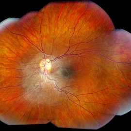
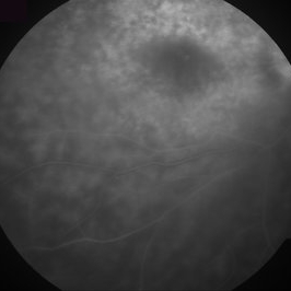
20-year-old patient with retinitis pigmentosa and pars planitis, recently s/p Retisert implant.
Photographer: William Yates, University of Colorado Eye Center
Imaging device: Optos camera (California model)
Condition/keywords: fluocinolone implant, pars planitis, retinitis pigmentosa
-
---thumb.jpg/image-square;max$300,300.ImageHandler) peripheral pre-retinal fibrosis and neovascularization with vitreous cutter
peripheral pre-retinal fibrosis and neovascularization with vitreous cutter
Feb 14 2013 by From the Collections of Thomas M. Aaberg, MD and Thomas M. Aaberg Jr., MD
intraoperative photo of peripheral pre-retinal fibrosis and neovascularization with vitreous cutter
Condition/keywords: pars planitis, peripheral retinal neovascularization
-
 Pars Planitis
-
 RP Retisert
Feb 2 2017 by Jeffrey L. Olson, MD
20-year-old patient with retinitis pigmentosa and pars planitis, recently s/p Retisert implant.
Photographer: William Yates, University of Colorado Eye Center
Imaging device: Optos California
Condition/keywords: pars planitis, retinitis pigmentosa
-
 peripheral pre-retinal fibrosis and neovascularization with vertical scissors
Feb 14 2013 by From the Collections of Thomas M. Aaberg, MD and Thomas M. Aaberg Jr., MD
intraoperative photo of peripheral pre-retinal fibrosis and neovascularization with vertical scissors
Condition/keywords: pars planitis, peripheral retinal neovascularization
-
 Epiretinal Membrane
Mar 27 2014 by Jason S. Calhoun
Patient with a history of pars planitis comes in with decreased vision in the left eye. VA is 20/30 in the left eye. Fundus photos show a rather large epiretinal membrane covering the optic nerve in the left eye.
Photographer: Jason S. Calhoun, Mayo Clinic Jacksonville, Department of Ophthalmology
Imaging device: TOPCON TRC 50-EX
Condition/keywords: epiretinal membrane (ERM), macular pucker
-
 RP Retisert
Feb 2 2017 by Jeffrey L. Olson, MD
20-year-old patient with retinitis pigmentosa and pars planitis, recently s/p Retisert implant
Photographer: William Yates, University of Colorado Eye Center
Imaging device: Optos California
Condition/keywords: retinitis pigmentosa (RP) dystrophy
-
---thumb.jpg/image-square;max$300,300.ImageHandler) Retinoschisis Secondary To Pars Planitis
Retinoschisis Secondary To Pars Planitis
Nov 26 2013 by Maurice F. Rabb
Retinoschisis secondary to pars planitis.
Condition/keywords: pars planitis, retinoschisis
-
 Pars Planitis
-
 Pars Planitis
-
 Pars Planitis
-
 Pars Planitis
 Loading…