Search results (135 results)
-
 Cyst of the Pars Plana
Nov 9 2012 by Norman Byer
This is a cyst of the pars plana located just anterior to the ora serrata in the lower temporal quadrant. It illustrates how far anterior one may visualize the fundus with indirect ophthalmoscopy and scleral indentation. Pars plana cysts are common lesions of no particular clinical significance.
Condition/keywords: cyst of the pars plana, lower temporal quadrant, ora serrata, scleral indentation
-
 Meridional Fold
Nov 9 2012 by Norman Byer
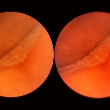
The next two photographs are of the same lesion in a 28-year-old woman. This view shows a sloping retinal mound with a radial retinal fold in the center. This is not a typical meridional fold for it stops short of the ora serrata and there is no dentate process. The upper temporal ora serrata and pars plana are well shown and peripheral cystoid degeneration is present posterior to the ora.
Condition/keywords: ora serrata, pars plana, peripheral cystoid degeneration, radial retinal fold, sloping retinal mound
-
 Retinal Detachment Right Eye Optomap
Mar 31 2014 by James B. Soque, CRA, OCT-C, COA, FOPS
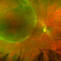
36-year-old white male presented with non traumatic retinal detachment OD, with six very distinct demarcation lines and isolated tear, and detachment parameters. Patient underwent PPV OD on 12/3/13 with 20% SF6 gas placement and face down in his first 1 month post op period.
Photographer: James Soque, CRA, COA
Imaging device: Optos Daytona
Condition/keywords: Cryopexy, demarcation line, gas pneumatic displacement, Optomap, Optos, pars plana vitrectomy (PPV), retinal tear, scanning laser ophthalmoscope
-
 Pars Plana Snowbank
Oct 9 2012 by Jeffrey G. Gross, MD, FASRS
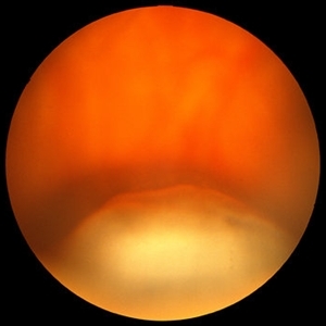
Pars plana snowbank.
Condition/keywords: pars plana, snowbank
-
 "Internal Mirroring" Effect by Intraocular Gas
Mar 25 2014 by Homayoun Tabandeh, MD, FASRS
"Internal mirroring" by residual intraocular gas in a highly myopic patient 3 weeks post repair of retinal detachment with pars plana vitrectomy and C3F8 gas.
Photographer: Danny Rivas
Condition/keywords: high myopia, intraocular gas
-
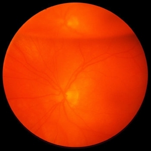
---thumb.jpg/image-square;max$300,300.ImageHandler) C3F8 gas bubble after retinal detachment surgery
C3F8 gas bubble after retinal detachment surgery
Feb 1 2013 by Sharon Fekrat, MD FACS FASRS
63 year old man s/p encircling scleral buckle and 23g pars plana vitrectomy for a macula off phakic rhegmatogenous retinal detachment. This fundus photograph shows the effect of the encircling buckle and the residual C3F8 intravitreal gas bubble in the right eye.
Photographer: Tiffanie Keaton, Duke Eye Imaging, Duke University Eye Center, Durham, NC
Imaging device: Optos
Condition/keywords: intravitreal gas bubble, vitrectomy
-
 Enclosed Ora Bay On The Temporal Side
Nov 9 2012 by Norman Byer
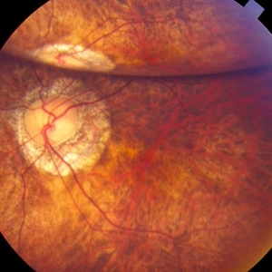
This is a developmental abnormality in a 59-year-old man. It is an enclosed ora bay on the temporal side, an isolated island of normal pars plana epithelium. It is important not to confuse this entity with a retinal break. It has smooth, sloping borders not a sharp, thin, visible retinal edge as a retinal break would have. The border looks exactly like that of the ora serrata, and the grayish pigmented base has the same appearance as the normal pars plana.
Condition/keywords: developmental abnormality, enclosed ora bay, grayish pigmented base, horizontal nasal meridian, pars plana epithelium, smooth sloping borders, temporal retina
-
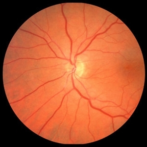
 Epiretinal Membrane
Oct 11 2012 by Michael P. Kelly, FOPS
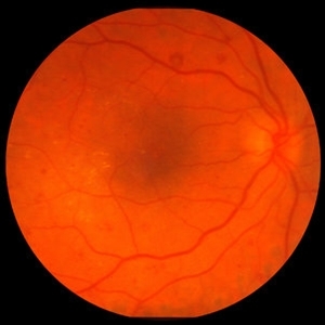
This is a patient with idiopathic panuveitis who developed a visually significant epiretinal membrane. Pars plana vitrectomy with membrane peeling was performed to remove the epiretinal proliferation. I recommend magnifying the image to see the exquisite detail centrally.
Photographer: Michael P. Kelly, FOPS Director, Duke Eye Center Labs, Duke Universtiy Hospital
Imaging device: Zeiss 450Plus
Condition/keywords: epiretinal membrane (ERM), panuveitis
-
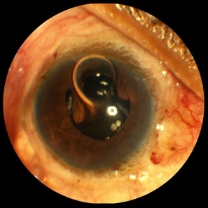 Gas Bubble Extending into Anterior Chamber
Oct 12 2012 by Jeffrey G. Gross, MD, FASRS
Gas bubble extending into anterior chamber in aphakic eye, after PPV.
Condition/keywords: anterior chamber, aphakic eye, gas bubble, pars plana vitrectomy (PPV)
-
 Senile Retinoschisis
Nov 9 2012 by Norman Byer
This is the same case as seen in the previous photograph but is a different view with the scleral indentation moved more anterior. The retinoschisis is seen to be very peripheral coming at least grossly right up to the ora serrata. Please notice how clear a view one can get of the ora serrata and pars plana using indirect ophthalmoscopy with scleral indentation.
Condition/keywords: ora serrata, retinoschisis, scleral indentation
-
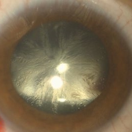 Lens Feathering
Nov 9 2016 by Nimrod Dar
26-year-old man with lens feathering a day after a pars plana vitrectomy for rhegmatogenous retinal detachment.
Photographer: Nimrod Dar, M.D, Meir Ophthalmology Department
Condition/keywords: lens feathering, vitrectomy
-
 Retinoschisis/Retinal Detachment
Oct 16 2012 by Jeffrey G. Gross, MD, FASRS
Retinoschisis/RD s/p PPV and laser.
Condition/keywords: laser, pars plana vitrectomy (PPV), retinoschisis
-
 Full-thickness Macular Hole
Aug 28 2012 by Sharon Fekrat, MD FACS FASRS
65 year old woman with a recurrent full thickness macular hole following previous 20 g pars plana vitrectomy in the right eye as well as an iatrogenic retinal hole in the papillomacular bundle. Both retinal defects are captured here in this Zeiss Stratus OCT image.
Photographer: Michael P. Kelly, FOPS Director, Duke Eye Labs, Duke University Eye Center, Durham, NC
Imaging device: Zeiss Cirrus
Condition/keywords: retinal break
-
---thumb.JPG/image-square;max$300,300.ImageHandler) Black Sunburst
Black Sunburst
Nov 25 2012 by Mallika Goyal, MD
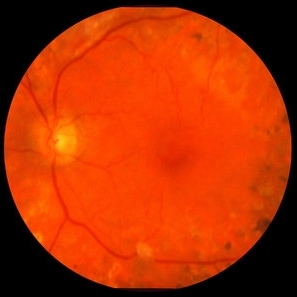
Left eye of a 19-year-old boy with sickle cell disease shows large salmon patch (decolourised subretinal heme and intraretinal heme) 3 days after pars plana vitrectomy for vitreous haemorrhage.
Photographer: Mallika Goyal, MD, Apollo Health City, Hyderabad, India
Condition/keywords: sickle cell
-
 Intravitreal Gas Bubble after PPV
Oct 12 2012 by Jeffrey G. Gross, MD, FASRS
Intravitreal gas bubble after PPV, with mirroring reflection of ON.
Condition/keywords: intravitreal gas bubble, mirroring reflection of ON, pars plana vitrectomy (PPV)
-
 Diamond Dust from Retinal Scraper
Oct 15 2012 by Jeffrey G. Gross, MD, FASRS
Diamond dust from retinal scraper, s/p PPV.
Condition/keywords: diamond dust, pars plana vitrectomy (PPV), retinal scraper
-
 Proliferative Diabetic Retinopathy with Severe Subhyaloid Hemorrhage
Oct 15 2012 by Jeffrey G. Gross, MD, FASRS
PDR with severe subhyaloid hemorrhage post-op, PPV.
Condition/keywords: pars plana vitrectomy (PPV), post-op, subhyaloid hemorrhage
-
---thumb.JPG/image-square;max$300,300.ImageHandler) Salmon Patch
Salmon Patch
Nov 25 2012 by Mallika Goyal, MD
Left eye of a 19-year-old boy with sickle cell disease shows large salmon patch (decolourised subretinal heme and intraretinal heme) 3 days after pars plana vitrectomy for vitreous haemorrhage.
Photographer: Mallika Goyal, MD, Apollo Health City, Hyderabad, India
Condition/keywords: sickle cell
-
 Vitreous Amyloidosis Slit Lamp Photo
Oct 23 2019 by Alexander D Port, MD
Slit lamp photograph preoperatively demonstrating dense symptomatic vitreous opacity in the setting of amyloidosis. The patient elected to undergo pars plana vitrectomy.
Condition/keywords: slit lamp photo, vitreous amyloidosis
-
 Proliferative Diabetic Retinopathy with Macular Traction
Oct 15 2012 by Jeffrey G. Gross, MD, FASRS
PDR with macular traction, post-op, PPV and PRP.
Condition/keywords: macular traction, pan-retinal photocoagulation (PRP), pars plana vitrectomy (PPV), post-op
-
---thumb.JPG/image-square;max$300,300.ImageHandler) Salmon Patch
Salmon Patch
Nov 25 2012 by Mallika Goyal, MD
Left eye of a 19-year-old boy with sickle cell disease shows large salmon patch (decolourised subretinal heme and intraretinal heme) 3 days after pars plana vitrectomy for vitreous haemorrhage.
Photographer: Mallika Goyal, MD, Apollo Health City, Hyderabad, India
Condition/keywords: sickle cell
-
 360 Degree Retinectomy
Sep 11 2020 by Sham Talati, DOMS
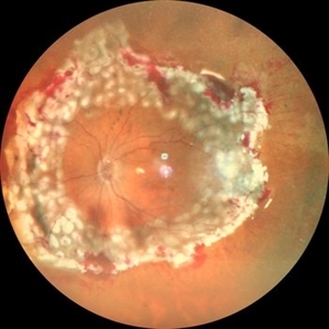
A case of retinal detachment with PVR. Patient underwent pars plana vitrectomy with silicon oil injection with 360 degree retinectomy.
Photographer: Dr. Sham Talati,Retina Foundation,Ahmedabad
Imaging device: Nidek Mirante
Condition/keywords: proliferative vitreoretinopathy (PVR), retinectomy
-
 Tools & Techniques for 23, 25, and 27G PPV
Dec 10 2012 by Yale L. Fisher, MD
Dr. Steve Charles reviews the tools and techniques he uses for 23G, 25G, and 27G PPV. NOTE: A narration by Dr. Steve Charles will soon be available for this movie- please check back periodically.
Condition/keywords: pars plana vitrectomy (PPV), video
-
---thumb.JPG/image-square;max$300,300.ImageHandler) Salmon Patch
Salmon Patch
Nov 25 2012 by Mallika Goyal, MD
Left eye of a 19-year-old boy with sickle cell disease shows large salmon patch (decolourised subretinal heme and intraretinal heme) 3 days after pars plana vitrectomy for vitreous haemorrhage.
Photographer: Mallika Goyal, MD, Apollo Health City, Hyderabad, India
Condition/keywords: sickle cell
 Loading…