Search results (9 results)
-
 Outer-Retinal-Tubulation
Jun 27 2013 by Jason S. Calhoun
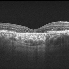
Patient with a history of wet macular degeneration and glaucoma in both eyes. VA is 20/50, right eye, 20/80, left eye. Patient is treated with Eylea in both eyes. Enhanced depth imaging OCT reveals a small like form of a cyst which in fact isn't a cyst at all. This is called outer retinal tubulation in which degenerating photo-receptors may become arranged in a circular or ovoid fashion. This is sometimes misdiagnosed as cystic changes in the retinal pigment epithelium or sub-retinal fluid.
Photographer: Jason S. Calhoun, Mayo Clinic Jacksonville, Florida
Imaging device: ZEISS OCT CIRRUS
Condition/keywords: optical coherence tomography (OCT)
-
 Outer Retinal Tubulation
Mar 27 2018 by Dhaivat Shah
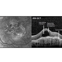
Outer retinal tubulation (ORT) is a feature of photoreceptor rearrangement after chronic retinal damage due to refractory cme, long standing CNVM or old trauma. Photoreceptors lose adhesions to surrounding structures, resulting in outward folding and formation of new lateral contact between photoreceptors to form round structure. They generally remains stable over time. It is important to recognize ORT on OCT because it indicates a refractory state of the pathological condition and poor visual prognosis, and likely not to benefit from any treatment. Here is a case of 62-year-old female with history of 4 previous anti-VEGF injection in left eye for CNVM, with the recent OCT showing formation of ORT with subfoveal scarred membrane.
Photographer: Dr Dhaivat Shah
Condition/keywords: choroidal neovascular membrane (CNVM), outer retinal tubulation
-
 Outer Retinal Tubulation
Jan 10 2015 by Thomas A. Ciulla, MD, MBA, FASRS
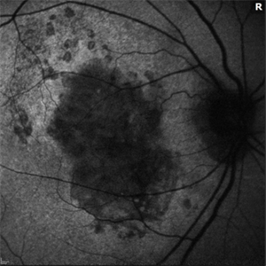
This 85-year-old man has a history of exudative AMD on the right and has undergone numerous anti-VEGF injections over the past 8 years. Visual acuity measured 20/60. OCT shows subretinal hyperreflective material with overlying outer retinal tubulation (round or oval structures with hyperreflective borders). Outer retinal tubulation is thought to result from invagination of photoreceptors at the junction of intact and atrophic outer retina. As in this case, greater lesion size, subretinal hyperreflective material and geographic atrophy are all associated with outer retinal tubulation. Poor visual function is also associated with outer retinal tubulation. Some reports suggest that outer retinal tubulation may be a predictor of enlargement of geographic atrophy.
Photographer: Stuart Alfred
Condition/keywords: outer retinal tubulation
-
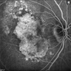
 Outer Retinal Tubulation
Jan 10 2015 by Thomas A. Ciulla, MD, MBA, FASRS
Autofluorescence images reveal geographic atrophy .
Photographer: Stuart Alfred
Condition/keywords: outer retinal tubulation
-
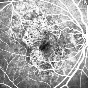
 Outer Retinal Tubulation
Jan 10 2015 by Thomas A. Ciulla, MD, MBA, FASRS
Fluorescein angiogram images reveal geographic atrophy and no active CNVM currently.
Photographer: Stuart Alfred
Condition/keywords: outer retinal tubulation
-
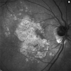 Outer Retinal Tubulation
Jan 10 2015 by Thomas A. Ciulla, MD, MBA, FASRS
Infrared images reveal geographic atrophy.
Photographer: Stuart Alfred
Condition/keywords: outer retinal tubulation
-
 Outer Retinal Tubulation
Mar 28 2018 by Rania G Estawro, FRCS
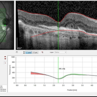
Enface OCT and line scan of outer retinal tubulation in a case of scarred CNV post multiple anti-VEGF injection.
Photographer: Rania Estawro, Al Watany Eye Hospital, Cairo, Egypt.
Imaging device: RTVue XR; Optovue, Fremont, CA, USA
Condition/keywords: outer retinal tubulation
-
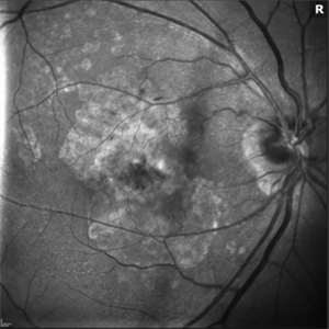 Outer Retinal Tubulation
Jan 10 2015 by Thomas A. Ciulla, MD, MBA, FASRS
Red-free images reveal geographic atrophy.
Photographer: Stuart Alfred
Condition/keywords: outer retinal tubulation
-
 Outer Retinal Tubulation
Jan 10 2015 by Thomas A. Ciulla, MD, MBA, FASRS
Fluorescein angiogram images reveal geographic atrophy and no active CNVM currently.
Photographer: Stuart Alfred
Condition/keywords: outer retinal tubulation
 Loading…