 Initializing download.
-
 By Thomas A. Ciulla, MD, MBA, FASRS
Indiana University School of Medicine - Uploaded on Jan 10, 2015.
- Last modified by Caroline Bozell on Jan 12, 2015.
- Rating
- Appears in
- Outer Retinal Tubulation
- Condition/keywords
- outer retinal tubulation
- Photographer
- Stuart Alfred
- Imaging device
- Scanning laser ophthalmoscope
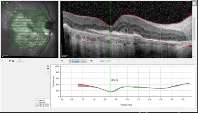
- Description
- This 85-year-old man has a history of exudative AMD on the right and has undergone numerous anti-VEGF injections over the past 8 years. Visual acuity measured 20/60. OCT shows subretinal hyperreflective material with overlying outer retinal tubulation (round or oval structures with hyperreflective borders). Outer retinal tubulation is thought to result from invagination of photoreceptors at the junction of intact and atrophic outer retina. As in this case, greater lesion size, subretinal hyperreflective material and geographic atrophy are all associated with outer retinal tubulation. Poor visual function is also associated with outer retinal tubulation. Some reports suggest that outer retinal tubulation may be a predictor of enlargement of geographic atrophy.


