Search results (308 results)
-
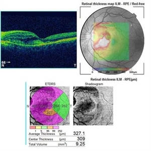
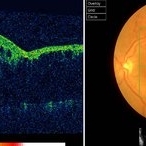
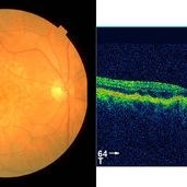
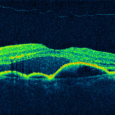
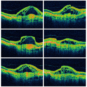 Wet Macular Degeneration OCT
Oct 13 2012 by Geoffrey G. Emerson, MD, PhD, FASRS
Condition/keywords: optical coherence tomography (OCT)
-
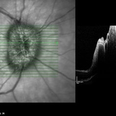
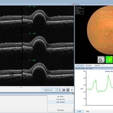 PED due to CSCR
Sep 2 2012 by Hamid Ahmadieh, MD
OCT image of a 37-year-old man with a serous PED secondary to CSCR.
Photographer: Hamid Ahmadieh, Ophthalmic Research Center, Labbafinejad Medical Center
Imaging device: Heidelberg Spectralis
Condition/keywords: central serous chorioretinopathy (CSCR), optical coherence tomography (OCT), pigment epithelial detachment (PED)
-
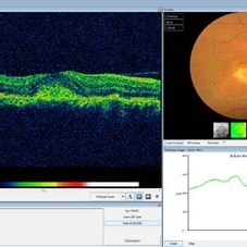
 Fibrovascular PED
Feb 21 2014 by Roy Schwartz, MD
72-year-old female with fibrovascular PED. Upper picture - PED with sub RPE hyper-reflective substance, in a multi-layered pattern, corresponding to fibrovascular PED. CME. Lower picture - PED flattened, a denser sub RPE hyperreflective substance is seen. CME resolved.
Condition/keywords: fibrovascular pigment epithelial detachment (PED), neovascular age-related macular degeneration (AMD), optical coherence tomography (OCT), ranibizumab
-
 Choroidal Osteoma Plus CNV
Sep 2 2012 by Hamid Ahmadieh, MD
Color fundus photograph and OCT imaging of a 47-year-old man with a juxtafoveal CNV superimposed on a choroidal osteoma.
Photographer: Hamid Ahmadieh, Ophthalmic Research Center, Labbafinejad Medical Center
Imaging device: Topcon
Condition/keywords: choroidal neovascularization (CNV), choroidal osteoma, optical coherence tomography (OCT)
-
 Chronic Active Central Serous Chorioretinopathy (CSCR)
Sep 11 2012 by Hamid Ahmadieh, MD
Color fundus photograph and OCT image of a 30-year-old man with chronic active CSCR.
Photographer: Hamid Ahmadieh, MD, Ophthalmic Research Center, Labbafinejad Medical Center, Shahid Beheshti University of Medical Sciences
Imaging device: Topcon
Condition/keywords: central serous chorioretinopathy (CSCR), optical coherence tomography (OCT)
-
 Whole Eye OCT
Jan 4 2019 by Netan Choudhry, MD, FRCS(C) FASRS
Swept-Source OCT montage of a 45-year-old male with Alports disease and posterior subcapsular cataract.
Photographer: John Golding BA, Vitreous Retina Macula Specialists of Toronto
Imaging device: Topcon DRI Triton
Condition/keywords: Alports disease, optical coherence tomography (OCT), swept source
-
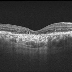
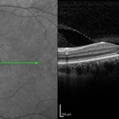
 OCT Myopic Staphyloma With Schisis and ERM
Apr 24 2014 by Scott E. Pautler, MD
OCT of high myope with asymptomatic macular schisis.
Imaging device: Heidelberg Spectralis
Condition/keywords: foveal schisis, maculopathy, maculoschisis, optical coherence tomography (OCT), pathologic myopia, staphyloma
-
 Angioid Streaks & CNV (Fig 5)
Sep 2 2012 by Hamid Ahmadieh, MD
OCT imaging of a 53-year-old woman with a juxtafoveal CNV secondary to angioid streaks.
Photographer: Hamid Ahmadieh, Ophthalmic Research Center, Labbafinejad Medical Center
Imaging device: Topcon
Condition/keywords: angioid streaks, choroidal neovascularization (CNV), optical coherence tomography (OCT)
-
 Macular Pseudohole
Jul 7 2015 by Hamid Ahmadieh, MD
Color fundus photograph and optical coherence tomography of the left eye of a 72-year-old woman with blurred vision due to epiretinal membrane. VA OS is 20/40 . Macular pseudohole is visible.
Photographer: Shabnam Poureh, Negah Eye Center, Tehran, Iran
Imaging device: Topcn
Condition/keywords: color fundus photograph, macular pseudohole, optical coherence tomography (OCT)
-
 Outer-Retinal-Tubulation
Jun 27 2013 by Jason S. Calhoun
Patient with a history of wet macular degeneration and glaucoma in both eyes. VA is 20/50, right eye, 20/80, left eye. Patient is treated with Eylea in both eyes. Enhanced depth imaging OCT reveals a small like form of a cyst which in fact isn't a cyst at all. This is called outer retinal tubulation in which degenerating photo-receptors may become arranged in a circular or ovoid fashion. This is sometimes misdiagnosed as cystic changes in the retinal pigment epithelium or sub-retinal fluid.
Photographer: Jason S. Calhoun, Mayo Clinic Jacksonville, Florida
Imaging device: ZEISS OCT CIRRUS
Condition/keywords: optical coherence tomography (OCT)
-
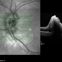
---thumb.jpg/image-square;max$300,300.ImageHandler) Optic Disc Drusen
Optic Disc Drusen
Jul 10 2013 by Hamid Ahmadieh, MD
SD-OCT image of the left eye of a 24-year-old woman with optic disc drusen and VA 20/20.
Photographer: Solmaz Shahmohammadi, Negah Eye Center, Tehran
Imaging device: Heidelberg Spectralis
Condition/keywords: optic disc drusen, optical coherence tomography (OCT)
-
 Branch Retinal Vein Occlusion (BRVO)
Sep 11 2012 by Hamid Ahmadieh, MD
Color fundus photograph and OCT image of a 60-year-old woman with a recent onset BRVO.
Photographer: Hamid Ahmadieh, MD, Ophthalmic Research Center, Labbafinejad Medical Center, Shahid Beheshti University of Medical Sciences
Imaging device: Topcon
Condition/keywords: branch retinal vein occlusion (BRVO), optical coherence tomography (OCT)
-
 Cone-Rod Dystrophy
Mar 15 2017 by Hamid Ahmadieh, MD
Infrared and OCT images of the left eye of a 16-year-old boy with decreased visual acuity and color vision deficiency due to cone-rod dystrophy.
Photographer: Abazarnezhad , Negah Eye Center, Tehran, Iran
Imaging device: Spectralis OCT
Condition/keywords: cone dystrophy, infrared image, optical coherence tomography (OCT)
-
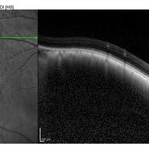 OCT of Large Choroidal Nevus
Jul 9 2014 by Susanna S. Park, MD, PhD
EDI OCT imaging of a large pigmented choridal nevus showing mild elevation with no associated subretinal fluid.
Photographer: Ellen Redenbo
Condition/keywords: choroidal nevus, optical coherence tomography (OCT)
-
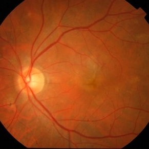 Active CNVM
Jul 11 2016 by Manish Nagpal, MD, FRCS (UK), FASRS
Colour photo showing an active CNVM.
Photographer: pooja barot
Condition/keywords: choroidal neovascular membrane (CNVM), optical coherence tomography (OCT)
-
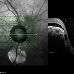
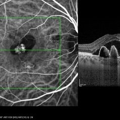
 Papilledema
Sep 8 2012 by Hamid Ahmadieh, MD
OCT of the optic nerve head of the right eye of a 55-year-old woman with a malignant intracranial tumor.
Photographer: Hamid Ahmadieh, MD, Ophthalmic Research Center, Labbafinejad Medical Center, Shahid Beheshti University of Medical Sciences
Imaging device: Heidelberg Spectralis
Condition/keywords: malignant intracranial tumor, optical coherence tomography (OCT), papilledema
-
 Coloboma of Disc & Choroid
Oct 6 2012 by Hamid Ahmadieh, MD
OCT image of a 25-year-old woman with serous retinal detachment secondary to coloboma of disc associated with coloboma of choroid.
Photographer: Hamid Ahmadieh, MD, Ophthalmic Research Center, Labbafinejad Medical Center, Shahid Beheshti University of Medical Sciences
Imaging device: Heidelberg Spectralis
Condition/keywords: coloboma of choroid, coloboma of optic disc, optical coherence tomography (OCT), serous retinal detachment
-
 Coloboma of Disc & Choroid
Oct 6 2012 by Hamid Ahmadieh, MD
OCT image of a 25-year-old woman with a small coloboma of choroid associated with coloboma of disc.
Photographer: Hamid Ahmadieh, MD, Ophthalmic Research Center, Labbafinejad Medical Center, Shahid Beheshti University of Medical Sciences
Imaging device: Heidelberg Spectralis
Condition/keywords: coloboma of choroid, coloboma of optic disc, optical coherence tomography (OCT)
-
 Dry Type AMD
Sep 22 2012 by Hamid Ahmadieh, MD
Color fundus photograph and OCT image of a 70-year-old woman with dry type AMD.
Photographer: Hamid Ahmadieh, MD, Ophthalmic Research Center, Labbafinejad Medical Center, Shahid Beheshti University of Medical Sciences
Imaging device: Topcon Fundus Camera & Topcon OCT
Condition/keywords: dry age-related macular degeneration (dry AMD), optical coherence tomography (OCT)
-
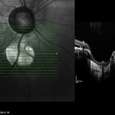
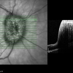
 Papilledema
Sep 8 2012 by Hamid Ahmadieh, MD
OCT of the optic nerve head of the left eye of a 55-year-old woman with a malignant intracranial tumor.
Photographer: Hamid Ahmadieh, MD, Ophthalmic Research Center, Labbafinejad Medical Center, Shahid Beheshti University of Medical Sciences
Imaging device: Heidelberg Spectralis
Condition/keywords: malignant intracranial tumor, optical coherence tomography (OCT), papilledema
-
 Polypoidal Choroidal Vasculopathy (ICG + OCT EDI)
May 16 2014 by Avris Romario Diparaja Siahaan
ICG + OCT (with EDI) (simultaneously)of a 46-year-old man with polypoidal choroidal vasculopathy (PCV).
Photographer: Avris Romario Diparaja Siahaan, Klinik Mata Nusantara
Imaging device: Heidelberg HRA + OCT Spectralis
Condition/keywords: optical coherence tomography (OCT), polypoidal choroidal vasculopathy (PCV)
-
 Papilledema
Sep 8 2012 by Hamid Ahmadieh, MD
OCT of the optic nerve head of the right eye of a 55-year-old woman with a malignant intracranial tumor.
Photographer: Hamid Ahmadieh, MD, Ophthalmic Research Center, Labbafinejad Medical Center, Shahid Beheshti University of Medical Sciences
Imaging device: Heidelberg Spectralis
Condition/keywords: optical coherence tomography (OCT), papilledema
-
 Bulls eye retinopathy
Nov 20 2012 by Roy Schwartz, MD
75-YEAR-OLD FEMALE PRESENTS WITH BILATERAL GRADUAL VISUAL LOSS 6/30 Dx BE PSEUDOPHAKIA + PCO BE BULLS EYE MACULOPATHY PER FA VA IMPROVES TO 6/10 S/P YAG CAPSULOTOMY OCT - BE MACULAR SUBRETINAL FLUID NO HISTORY OF CHLOROQUINE THERAPY NO DRUSEN OR SIGNS OF AMD WORKING DIAGNOSIS - BE CHRONIC CSCR
Imaging device: Heidelberg spectralis
Condition/keywords: bull's eye maculopathy, optical coherence tomography (OCT)
-
 Choroidal Granuloma Secondary to Tuberculosis
Mar 14 2013 by Eduardo Torres-Porras, MD
OCT scan through the granuloma shows attachment of the retinal pigment epithelial-choriocapillaris layer and the neurosensory retina over the granuloma (“contact” sign), inflammatory retinal infiltrate in the deeper retinal layers and subretinal fluid.
Photographer: Eduardo Torres Porras, Laser y ultrasonido ocular de Puebla
Imaging device: Cirrus
Condition/keywords: optical coherence tomography (OCT), tubercular choroidal granuloma
-
 OCT Cirrus 5 Line Scan ARMD SRF RPED Stage 3 PVD
Mar 6 2013 by James B. Soque, CRA, OCT-C, COA, FOPS
Zeiss Cirrus OCT 4000, 5 Line Scan , 91-year-old white female with peripapillary SRN with subretinal heme, serous fluid, and a stage 3 PVD, still attached at the optic nerve.
Photographer: James Soque, CRA, COA, Island-Retina
Imaging device: Zeiss Cirrus 4000 SD OCT with 6.0.2.81 Software
Condition/keywords: optical coherence tomography (OCT)
 Loading…