Search results (98 results)
-
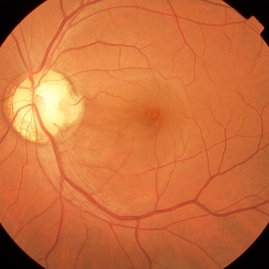 Pit Macular Syndrome
Mar 21 2013 by Yusuke Oshima, MD, PhD
Fundus photograph of a 38-year-old man with macular detachment associated with an optic disc pit.
Photographer: Yusuke Takada, Osaka University Graduate School of Medicine
Condition/keywords: congenital optic nerve pit
-
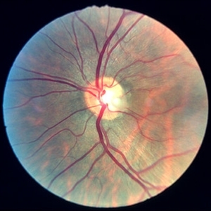
 Optic Nerve Pit v Coloboma
Feb 19 2013 by From the Collections of Thomas M. Aaberg, MD and Thomas M. Aaberg Jr., MD
Optic Nerve Pit v Coloboma, 20/20, right eye.
Condition/keywords: optic nerve coloboma, optic nerve pit
-
 Optic Pit
Oct 12 2012 by Gregg T. Kokame, MD, MMM, FASRS
Optic pit
Photographer: Jaclyn Pisano, Retina Consultants of Hawaii
Imaging device: Zeiss FF-450 plus
Condition/keywords: congenital optic nerve pit
-
 Optic Nerve Pit
Aug 30 2012 by Raj K. Maturi, MD
congenital optic nerve pit with chronic pigment changes in macula due to detachment
Photographer: Tom Steele, CRA, Midwest Eye Institute
Imaging device: Topcon Ex
Condition/keywords: optic nerve pit
-
 Optic Nerve Pit / Two in One Nerve
Feb 19 2013 by From the Collections of Thomas M. Aaberg, MD and Thomas M. Aaberg Jr., MD
Right of stereo; 20/20; superior temporal arcuate defect.
Condition/keywords: optic nerve pit, two in one nerve
-
 Optic Nerve Pit
Aug 30 2012 by Raj K. Maturi, MD
Photographer: Tom Steele, CRA, Midwest Eye Institute
Imaging device: Topcon Ex
Condition/keywords: optic nerve pit
-
 Optic Nerve Pit OCT
Aug 24 2012 by John S. King, MD
8 yo c dec vision
Photographer: Kristin Konecki, OcuSight Eye Care Center, Rochester, NY
Condition/keywords: congenital optic nerve pit
-
 Optic Nerve Pit OD
Aug 24 2012 by John S. King, MD
8 yo with decreased vision
Photographer: Kristin Konecki, OcuSight Eye Care Center, Rochester, NY
Condition/keywords: congenital optic nerve pit
-
 Optic disc pit 3
Jan 11 2013 by Alex P. Hunyor, MD
Optic disc pit left eye.
Condition/keywords: congenital optic nerve pit, optic disc pit
-
 Optic Nerve Pit
Aug 30 2012 by Raj K. Maturi, MD
Photographer: Tom Steele, CRA, Midwest Eye Institute
Imaging device: Topcon Ex
Condition/keywords: optic nerve pit
-
 Bilateral Optic Nerve Pits - ON OCTs
Sep 18 2012 by Pauline T Merrill, MD, FASRS
Optic nerve OCTs of a 77-year-old woman with bilateral optic nerve pits and glaucoma, stable over 20 years.
Photographer: Karen Parque, Illinois Retina Associates, Chicago, IL
Imaging device: Zeiss Cirrus
Condition/keywords: optic nerve pit
-
 Optic Nerve Pit (Color Picture)
Sep 25 2013 by Alexandre Durao Alves Pereira, MD
Optic nerve pit (color picture).
Photographer: Alexandre Pereira
Condition/keywords: color photo, optic nerve pit
-
 Optic disc pit - R stereo
Jan 11 2013 by Alex P. Hunyor, MD
Optic disc pit - R stereo. Note chronic RPE changes from subretinal fluid
Condition/keywords: congenital optic nerve pit, optic disc pit
-
 Optic Nerve Pit
Aug 30 2012 by Raj K. Maturi, MD
Photographer: Tom Steele, CRA, Midwest Eye Institute
Imaging device: Topcon Ex
Condition/keywords: optic nerve pit
-
 Optic disc pit - L stereo
Jan 11 2013 by Alex P. Hunyor, MD
Optic disc pit - L stereo.
Condition/keywords: congenital optic nerve pit, optic disc pit
-
 Optic Nerve Pit
Aug 30 2012 by Raj K. Maturi, MD
Photographer: Tom Steele, CRA, Midwest Eye Institute
Imaging device: Topcon Ex
Condition/keywords: optic nerve pit
-
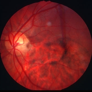
 Optic Nerve Pit v Coloboma / fellow eye
Feb 19 2013 by From the Collections of Thomas M. Aaberg, MD and Thomas M. Aaberg Jr., MD
Optic Nerve Pit v Coloboma / fellow eye, 20/25, left eye
Condition/keywords: optic nerve coloboma, optic nerve pit
-
 Optic Nerve Pit 2
Aug 24 2012 by John S. King, MD
8 yo c dec vision
Photographer: Kristin Konecki, OcuSight Eye Care Center, Rochester, NY
Condition/keywords: congenital optic nerve pit
-
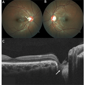 Optic Disc Pit
Nov 8 2021 by Michael Grinton
Optic disc pits are rare congenital abnormalities of the optic nerve head. Colour fundus image of an asymptomatic 18-year old male shows an optic disc pit in the right eye (A, white arrow); a small, grey, oval shaped excavation in the temporal segment of the optic disc. These pits are usually unilateral (B shows normal colour fundus of left eye) and asymptomatic. Imaging with optical coherence tomography (C) shows the optic disc pit in cross section (white arrow) and normal macular structure. In some patients with the condition, fluid can accumulate underneath the macular (serous macular detachment).
Condition/keywords: Optic disc pit, Optic nerve pit, Optic pit
-
 Optic Nerve Head Pit with Fluid
Feb 20 2013 by From the Collections of Thomas M. Aaberg, MD and Thomas M. Aaberg Jr., MD
Optic Nerve Head Pit Red Free Photo
Condition/keywords: optic nerve pit, red-free
-
 ON pits OD mac OCT
Sep 18 2012 by Pauline T Merrill, MD, FASRS
Right macular OCT of a 77-year-old woman with bilateral optic nerve pits and glaucoma, stable over 20 years.
Photographer: Karen Parque, Illinois Retina Associates, Chicago, IL
Imaging device: Zeiss Cirrus
-
 Optic Nerve Pit OD
Aug 24 2012 by John S. King, MD
8 yr old c decreased vision FA
Photographer: Kristin Konecki, OcuSight Eye Care Center, Rochester, NY
Condition/keywords: congenital optic nerve pit
-
 Optic Nerve Head Pit
Feb 20 2013 by From the Collections of Thomas M. Aaberg, MD and Thomas M. Aaberg Jr., MD
optic nerve head pit Red free photograph
Condition/keywords: optic nerve pit
-
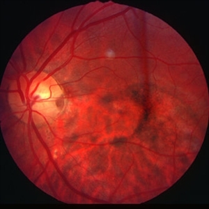
 Optic Disc Pit Maculopathy
Aug 20 2018 by DIEGO A BUESO PONCE, MD
Fundus photograph of an 19-year-old female with congenital optic disc pit and associated maculopathy with subretinal fluid and retinoschisis.
Photographer: Diego Bueso Ponce, Clinica Unidad Laser, Barranquilla Colombia
Imaging device: Topcon DRI OCT Triton, Swept source OCT
Condition/keywords: congenital optic nerve pit, maculopathy, optic disc pit, retinoschisis
-
 Partial Optic Disc Avulsion with Optic Disc Pit
Jul 1 2018 by John S. King, MD
16-year-old with acute loss of vision after blunt finger injury to eye while playing football. This photo is three weeks post-injury. Vision HM. Retinal striae with subhyaloid heme. Decreased retinal whitening. Peripapillary heme clearing, and temporal optic disc avulsion with optic disc pit can be seen.
Photographer: Maisee Yang
Imaging device: Topcon
Condition/keywords: epiretinal membrane (ERM), optic nerve head avulsion, optic nerve pit, traumatic optic neuropathy
 Loading…