Search results (152 results)
-
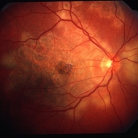
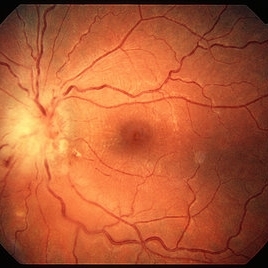 Toxoplasma Neuroretinitis (Jensen`s Disease)
Feb 25 2013 by Henry J. Kaplan, MD
Toxoplasma neuroretinitis in the left eye of a patient with macular star formation, retinitis adjacent to the optic nerve head with disc swelling.
Condition/keywords: Jensen disease, ocular toxoplasmosis, toxoplasmosis
-
 Optic Nerve Head Drusen With Idiopathic CNV
Feb 17 2017 by Kristen Wagner
22-year-old female fundus photograph of a right eye with Optic Nerve Drusen with Idiopathic CNV.
Photographer: Kristen Wagner, COT, OSC Ophthalmic Photographer, Tennessee Retina, Nashville TN
Condition/keywords: choroidal neovascularization (CNV), drusen of optic disc, optic disc drusen
-
 Gyrate Atrophy of Choroid and Retina
Apr 19 2014 by Mallika Goyal, MD
Right eye fundus of a 45-year-old male patient with advanced gyrate atrophy of the choroid and retina with macular sparing. Optic nerve head is healthy.
Photographer: Mallika Goyal, MD, Apollo Health City, Hyderabad, India
Condition/keywords: choroid, gyrate atrophy
-
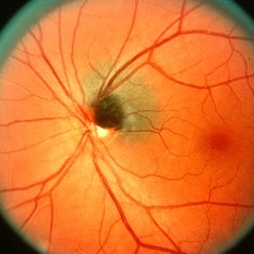
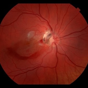
 Toxocara Granuloma
Feb 25 2013 by Henry J. Kaplan, MD
Toxocara granuloma of the optic nerve head.
Condition/keywords: ocular toxoplasmosis, toxocara granuloma, toxocariasis
-
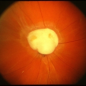
 Melanocytoma
May 2 2013 by Henry J. Kaplan, MD
Melanocytoma of the optic nerve head.
Condition/keywords: melanocytoma
-
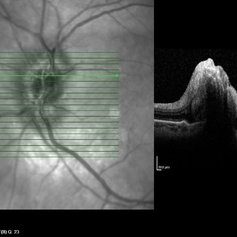
 Papilledema
Sep 8 2012 by Hamid Ahmadieh, MD
OCT of the optic nerve head of the right eye of a 55-year-old woman with a malignant intracranial tumor.
Photographer: Hamid Ahmadieh, MD, Ophthalmic Research Center, Labbafinejad Medical Center, Shahid Beheshti University of Medical Sciences
Imaging device: Heidelberg Spectralis
Condition/keywords: malignant intracranial tumor, optical coherence tomography (OCT), papilledema
-
---thumb.JPG/image-square;max$300,300.ImageHandler) SLE retinopathy
SLE retinopathy
Nov 18 2013 by Mallika Goyal, MD
Occlusive retinitis in a lady with SLE; optic nerve head pallor suggestive of prior optic neuritis or ischaemic optic neuropathy.
Photographer: Mallika Goyal, MD, Apollo Health City, Hyderabad
Condition/keywords: systemic lupus erythematosus (SLE) retinopathy
-
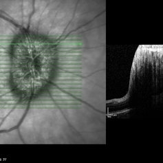
 Papilledema
Sep 8 2012 by Hamid Ahmadieh, MD
OCT of the optic nerve head of the left eye of a 55-year-old woman with a malignant intracranial tumor.
Photographer: Hamid Ahmadieh, MD, Ophthalmic Research Center, Labbafinejad Medical Center, Shahid Beheshti University of Medical Sciences
Imaging device: Heidelberg Spectralis
Condition/keywords: malignant intracranial tumor, optical coherence tomography (OCT), papilledema
-
 Papilledema
Sep 8 2012 by Hamid Ahmadieh, MD
OCT of the optic nerve head of the right eye of a 55-year-old woman with a malignant intracranial tumor.
Photographer: Hamid Ahmadieh, MD, Ophthalmic Research Center, Labbafinejad Medical Center, Shahid Beheshti University of Medical Sciences
Imaging device: Heidelberg Spectralis
Condition/keywords: optical coherence tomography (OCT), papilledema
-
 Remnant of Hyaloidal Artery
Feb 5 2014 by Gerardo Garcia-Aguirre, MD
Fundus photograph of the left eye of a 14-year-old asymptomatic female. The photograph is focused on the retina, and a prepapillary vitreous opacity is observed (white arrows). The opacity is attached to the origin of the retinal vessels in the optic nerve head.
Photographer: Gerardo Garcia-Aguirre, MD
Condition/keywords: persistence of the hyaloid artery
-
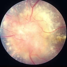
 Optic Nerve Head Drusen
Feb 12 2015 by Timothy S Fuller, MD
Fundus photograph of a 34-year-old woman with striking, asymptomatic optic nerve head drusen.
Photographer: Nice Hesse, Texas Retina Associates
Condition/keywords: drusen of optic disc
-
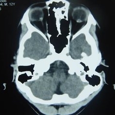 Orbital CT Scan in Optic Nerve Drusen
Mar 27 2013 by Henry J. Kaplan, MD
Axial CT scan of orbit demonstrates high density spot on optic nerve head on both sides #4.
Condition/keywords: drusen of optic disc, optic disc drusen, optic nerve drusen
-
 Remnant of Hyaloidal Artery
Feb 5 2014 by Gerardo Garcia-Aguirre, MD
Fundus photograph of the left eye of a 14-year-old asymptomatic female. The photograph is focused on the posterior vitreous where a prepapillary vitreous opacity is observed (white arrows). The opacity is attached to the origin of the retinal vessels in the optic nerve head.
Photographer: Gerardo Garcia-Aguirre, MD
Condition/keywords: persistence of the hyaloid artery
-
 Post Traumatic Optic Nerve Head Avulsion
Nov 18 2017 by Vishal Agrawal, MD, FRCS,FACS,FASRS
Right eye fundus picture of a 24-year-old male patient who suffered blunt trauma 7 days back with a wooden stick . He presented with NLP vision with a non reacting dilated pupil. Fundus montage picture shows ONH avulsion,CRAO,peripapillary resolving hemorrhages and cicatricial tissue at the edge.
Photographer: Vishal Agrawal, MD, SMS Medical College, Jaipur, India
Imaging device: Zeiss 524
Condition/keywords: avulsion, central retinal artery occlusion (CRAO)
-
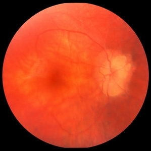
 Bilateral Optic Nerve Involvement in Sarcoidosis
Feb 25 2013 by Henry J. Kaplan, MD
Optic nerve head granuloma of sarcoidosis with severe infiltration and exudation in the left eye of the same patient #2.
Condition/keywords: bilateral involvement, sarcoid granuloma
-
 Serpiginous Choroiditis
Feb 25 2013 by Henry J. Kaplan, MD
Serpiginous choroiditis, right eye. Both active and inactive lesions clearly visible; active lesions are the yellowish subretinal area most prominant nasal to optic nerve head and also around the inferior arcade and temporal to the macular lesion.
Condition/keywords: serpiginous choroiditis
-
 Optic Nerve Head Drusen - Red Free
Oct 5 2013 by Roy Schwartz, MD
Optice nerve head drusen in a right eye, red-free image.
Photographer: Galit Yair-Pur
Condition/keywords: disc drusen, drusen of optic disc, optic nerve drusen, red-free
-
 Acute Traumatic Optic Nerve Avulsion
Feb 19 2016 by Mahdi Mwas
Fundus photograph of a 24-year-old gentleman, involved in a road traffic accident resulting in left no perception of light.
Photographer: Mahdi Mwas, FRCS, DRCOphth, Jordan
Condition/keywords: optic nerve head avulsion
-
 Remnant of Hyaloidal Artery
Feb 5 2014 by Gerardo Garcia-Aguirre, MD
Video of the fundus of the left eye of a 14-year-old asymptomatic female, where a prepapillary vitreous opacity is observed. The opacity is attached to the origin of the retinal vessels in the optic nerve head, and is considered to be a remnant of the hyaloidal artery.
Photographer: Gerardo Garcia-Aguirre, MD
Condition/keywords: persistence of the hyaloid artery
-
 Optic Nerve Head Drusen
Oct 16 2012 by Jeffrey G. Gross, MD, FASRS
Optic nerve head drusen.
Condition/keywords: optic nerve drusen
-
 Partial Optic Disc Avulsion with Optic Disc Pit
Jul 1 2018 by John S. King, MD
16-year-old with acute loss of vision after blunt finger injury to eye while playing football. This photo is three weeks post-injury. Vision HM.
Photographer: Maisee Yang
Imaging device: Topcon
Condition/keywords: epiretinal membrane (ERM), optic disc pit, optic nerve head avulsion, traumatic optic neuropathy
-
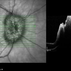
 cRORA
cRORA
Aug 5 2020 by Dhaivat Shah
A 54-year-old healthy male presented to us with a decreased vision in right eye since past 8 years. The patient gave a history of bleed in right eye before 8 years for which some intravitreal injection was given; post which there no major visual improvement. No details or documentation was available regarding the same. His BCVA in the right eye was 5/60. Fundus examination revealed a sharply demarcated hypopigmented patch over the macula with mild posterior excavation suggestive of macular scar. OCT image shows foveal thinning with loss of Retinal pigment epithelium and outer retinal layers (RORA). There are 2 types of RORAs, complete and incomplete. Complete RORA and incomplete RORA are entities defined by various imaging modalities describing atrophy of the retinal pigment epithelial and the outer retinal layers. OCT imaging defines incomplete RORA (iRORA) as a region of signal hyper transmission into the choroid and a corresponding zone of attenuation ordisruption of the RPE (<250um) and evidence of overlying photoreceptor degeneration (<250um). There should not be any RPE tear associated with it. OCT imaging describes complete RORA (cRORA) based on 4 inclusion criteria. These include, area of hypertransmission of more than 250um, zone of attenuation or disruption of the RPE of more than 250um in diameter, evidence of overlying photoreceptor degeneration and absence of scrolled RPE or other signs of an RPE tear. Other modalities used to define these include fundus autoflourescence(FAF), near infrared reflectance(NIR) and color fundus photograph(CFP). On CFP, it shows a sharply demarcated hypopigmented of >250um size with better visibility of choroidal vessels. FAF shows a hypo autoflourescent patch with sharply demarcated borders of size >250um, the colour of which is similar to that of the optic nerve head or retinal blood vessels excluding any pigmentation or artefact. On NIR, it shows a hyperreflective area with sharply demarcated borders of >250um size excluding any artefact. RORA can be seen in conditions like geographical atrophy in ARMD, central areolar choroidal dystrophy, atrophy secondary to anti-VEGF treatment. References: 1. Sadda SR, Guymer R, Holz FG, et al. Consensus Definition for Atrophy Associated with Age-Related Macular Degeneration on OCT: Classification of Atrophy Report 3 [published correction appears in Ophthalmology. 2019 Jan;126(1):177]. Ophthalmology. 2018;125(4):537-548. 2. Guymer RH, Rosenfeld PJ, Curcio CA, et al. Incomplete Retinal Pigment Epithelial and Outer Retinal Atrophy in Age-Related Macular Degeneration: Classification of Atrophy Meeting Report 4. Ophthalmology. 2020;127(3):394-409. 3. Eng VA, Rayess N, Nguyen HV, Leng T. Complete RPE and outer retinal atrophy in patients receiving anti-VEGF treatment for neovascular age-related macular degeneration. PLoS One. 2020;15(5):e0232353.
Photographer: Miss Anjum Zafar Khan
Imaging device: Choithram Netralaya
Condition/keywords: macular scar, outer retina, retinal pigment epithelium
-
 Optic Nerve Head Drusen - Fundus Image
Oct 5 2013 by Roy Schwartz, MD
Optice nerve head drusen in a right eye, fundus image.
Photographer: Galit Yair-Pur
Condition/keywords: disc drusen, drusen of optic disc, optic nerve drusen, red-free
-
 Remnant of Hyaloidal Artery
Feb 5 2014 by Gerardo Garcia-Aguirre, MD
Fundus photograph of the left eye of a 14-year-old asymptomatic female. The photograph is focused on the posterior vitreous where a prepapillary vitreous opacity is observed (see next picture where opacity is marked by arrows). The opacity is attached to the origin of the retinal vessels in the optic nerve head.
Photographer: Gerardo Garcia-Aguirre, MD
Condition/keywords: persistence of the hyaloid artery
-
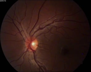
 Anterior ischemic optic neuropathy slide 1
Oct 22 2012 by Ronald C. Gentile, MD
70-year-old women with acute loss of vision in the left eye. Review of symptoms was significant for temporal arteritis and ESR was very high. Fundus examination of the left eye had a swollen white optic nerve head with a few peri-papillary cotton wool spots.
Photographer: The New York Eye & Ear Infirmary Department of Medical Imaging
Condition/keywords: anterior ischemic optic neuropathy, choroidal ischemia, temporal arteritis
 Loading…