Search results (69 results)
-
 Optic disc pit 2
-
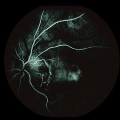
 Optic Pit FA
Jul 4 2012 by John T. Thompson, MD
Hyperfluorescence in optic pit due to fluorescein leakage
Imaging device: Zeiss FF4
Condition/keywords: fluorescein leakage, optic disc pit
-
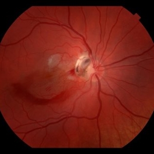
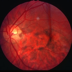
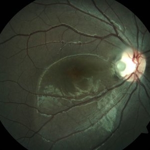
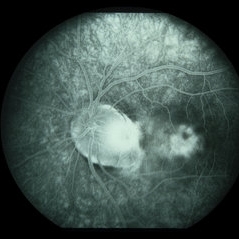
 Pit Macular Syndrome
Mar 21 2013 by Yusuke Oshima, MD, PhD
Fundus photograph of a 38-year-old man with macular detachment associated with an optic disc pit.
Photographer: Yusuke Takada, Osaka University Graduate School of Medicine
Condition/keywords: congenital optic nerve pit
-
 Partial Optic Disc Avulsion with Optic Disc Pit
Jul 1 2018 by John S. King, MD
16-year-old with acute loss of vision after blunt finger injury to eye while playing football. This photo is three weeks post-injury. Vision HM.
Photographer: Maisee Yang
Imaging device: Topcon
Condition/keywords: epiretinal membrane (ERM), optic disc pit, optic nerve head avulsion, traumatic optic neuropathy
-
 Optic Pit With Old Resolved Subretinal Fluid
Feb 20 2013 by From the Collections of Thomas M. Aaberg, MD and Thomas M. Aaberg Jr., MD
20/200.
Condition/keywords: optic disc pit, resolved subretinal fluid
-
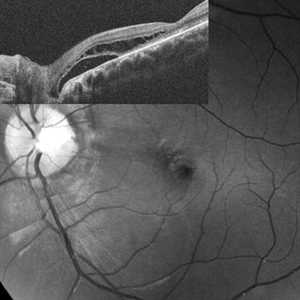 Optic Disc Pit Schisis RD
Apr 29 2013 by Michael Colucciello, MD, FASRS
Optic disc pit with peripapillary RD and macular schisis, fundus photograph and SD-OCT overlay.
Condition/keywords: optic disc pit
-
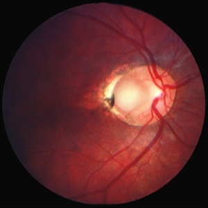
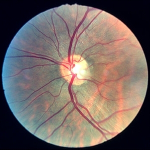 Optic disc pit 3
Jan 11 2013 by Alex P. Hunyor, MD
Optic disc pit left eye.
Condition/keywords: congenital optic nerve pit, optic disc pit
-
 Optic Pit With Subretinal Fluid and Macular Cyst
Feb 20 2013 by From the Collections of Thomas M. Aaberg, MD and Thomas M. Aaberg Jr., MD
No history.
Condition/keywords: optic disc pit, subretinal fluid
-
 Optic disc pit - R stereo
Jan 11 2013 by Alex P. Hunyor, MD
Optic disc pit - R stereo. Note chronic RPE changes from subretinal fluid
Condition/keywords: congenital optic nerve pit, optic disc pit
-
 Optic disc pit - L stereo
Jan 11 2013 by Alex P. Hunyor, MD
Optic disc pit - L stereo.
Condition/keywords: congenital optic nerve pit, optic disc pit
-
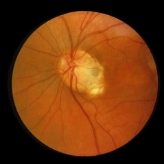
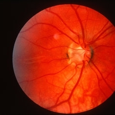 Optic pit
May 2 2013 by Henry J. Kaplan, MD
Optic pit in the inferotemporal part of the optic disc.
Condition/keywords: optic disc pit
-
 Optic Pit Red Free Photo
Jan 9 2014 by Susanna S. Park, MD, PhD
Red-free fundus photograph of a young man with recent vision loss from maculopathy associated with optic disc pit. Macular schisis and detachment with outer lamellar hole was noted preoperatively
Photographer: Ellen Redenbo, University of California Davis
Imaging device: Topcon
Condition/keywords: lamellar macular hole, macular schisis, optic disc pit, subretinal fluid
-
 Optic Disc Pit
Jun 3 2014 by Neha Goel, MS DNB FRCS (Glasg)
Fundus photograph of the right eye of a 15-year-old male.
Photographer: Neha Goel
Imaging device: Zeiss Visucam
Condition/keywords: optic disc pit
-
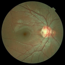
 Optic Disc Pit
Jun 4 2014 by Henry J. Kaplan, MD
Optic disc pit in the temporal part of optic nerve with associated CSR.
Condition/keywords: central serous retinopathy (CSR), optic disc pit
-
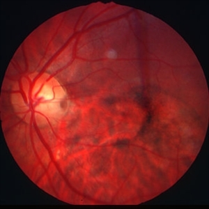
 Optic Disc Pit Maculopathy
Aug 20 2018 by DIEGO A BUESO PONCE, MD
Fundus photograph of an 19-year-old female with congenital optic disc pit and associated maculopathy with subretinal fluid and retinoschisis.
Photographer: Diego Bueso Ponce, Clinica Unidad Laser, Barranquilla Colombia
Imaging device: Topcon DRI OCT Triton, Swept source OCT
Condition/keywords: congenital optic nerve pit, maculopathy, optic disc pit, retinoschisis
-
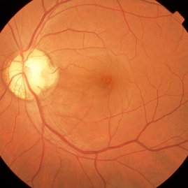
 Optic Disc Pit
Nov 27 2016 by Rita Couceiro, MD, MS
15-year-old boy with an optic disc pit of the right eye (incidental finding during routine fundoscopy).
Photographer: Andreia Rocha
Condition/keywords: optic disc pit
-
 Partial Optic Disc Avulsion with Optic Disc Pit
Jul 1 2018 by John S. King, MD
16-year-old with acute loss of vision after blunt finger injury to eye while playing football. This photo is three weeks post-injury. Vision HM. Retinal striae with subhyaloid heme. Decreased retinal whitening. Peripapillary heme clearing, and temporal optic disc avulsion with optic disc pit can be seen.
Photographer: Maisee Yang
Imaging device: Topcon
Condition/keywords: epiretinal membrane (ERM), optic nerve head avulsion, optic nerve pit, traumatic optic neuropathy
-
 Coloboma
Sep 7 2018 by John S. King, MD
11-year-old white female with bilateral optic nerve and retinochoroidal colobomas and an optic nerve pit in the right eye looking almost like pseudoduplication of the optic nerve. She is currently 20/30 OD and 20/20 OS. She has a history of laser by Dr. Zocchi about 10 years ago for a low lying, macula involving, serous retinal detachment, and has responded well.
Photographer: Stacey Coleman
Imaging device: Topcon
Condition/keywords: chorioretinal coloboma, inferior optic nerve coloboma, optic disc pit
-
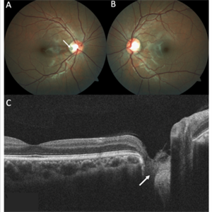 Optic Disc Pit
Nov 8 2021 by Michael Grinton
Optic disc pits are rare congenital abnormalities of the optic nerve head. Colour fundus image of an asymptomatic 18-year old male shows an optic disc pit in the right eye (A, white arrow); a small, grey, oval shaped excavation in the temporal segment of the optic disc. These pits are usually unilateral (B shows normal colour fundus of left eye) and asymptomatic. Imaging with optical coherence tomography (C) shows the optic disc pit in cross section (white arrow) and normal macular structure. In some patients with the condition, fluid can accumulate underneath the macular (serous macular detachment).
Condition/keywords: Optic disc pit, Optic nerve pit, Optic pit
-
 Vertical OCT Scan Through Right Optic Disc Pit
Jul 20 2019 by Arwa Azmeh, MD, PhD
Fundus photograph of 38-year-old healthy man with right optic disc pit, who recently noticed slightly blurred vision in right eye while closing the left eye. BCVA was 20/25 in OD and 20/20 in OS. IOP was 15mmHg OD and 14 mmHg OS. Right fundus exam showed small optic disc pit near the temporal rim of optic disc with abnormal reflex of nasal macula. Left fundus was normal. Late FA of right optic disc showed no leakage or staining of optic disc. Macular OCT showed normal foveal contour with no subretinal fluid or macular edema. There was significant reduction in RNFL thickness in the temporal sector in right eye. Coloboma is clearly seen on vertical OCT scan as well as horizontal scans through right optic pit.
Photographer: Ebtisam Aljbeili, Damascus university, Almouassat university hospital
Imaging device: Heidelberg Spectralis 2
Condition/keywords: optic pit, optical coherence tomography (OCT)
-
 Partial Optic Disc Avulsion with Optic Disc Pit
Jul 1 2018 by John S. King, MD
16-year-old with acute loss of vision after blunt finger injury to eye while playing football. Five days post-injury. Vision HM. Decreasing heme and retinal whitening.
Imaging device: Optos
Condition/keywords: traumatic optic neuropathy
-
 Optic disc Pit and CSR
Jun 4 2014 by Henry J. Kaplan, MD
Arterial phase F/A of the same patient shows hypofluorescence of the pit area and hyperfluorescence secondary to pooling in the serous detachment part. #2
Condition/keywords: central serous retinopathy (CSR), optic disc pit
-
 Optic Disc Pit and CSR
Jun 4 2014 by Henry J. Kaplan, MD
Late phase angiogram clearly shows the abnormally large optic nerve with temporal pit which is stained. #4
Condition/keywords: central serous retinopathy (CSR), optic disc pit
-
 Horizontal OCT Scan Through Right Optic Pit
Jul 20 2019 by Arwa Azmeh, MD, PhD
Fundus photograph of 38-year-old healthy man with right optic disc pit, who recently noticed slightly blurred vision in right eye while closing the left eye. BCVA was 20/25 in OD and 20/20 in OS. IOP was 15mmHg OD and 14 mmHg OS. Right fundus exam showed small optic disc pit near the temporal rim of optic disc with abnormal reflex of nasal macula. Left fundus was normal. Late FA of right optic disc showed no leakage or staining of optic disc. Macular OCT showed normal foveal contour with no subretinal fluid or macular edema. There was significant reduction in RNFL thickness in the temporal sector in right eye. Coloboma is clearly seen on vertical OCT scan as well as horizontal scans through right optic pit.
Photographer: Ebtisam Aljbeili, Damascus university, Almouassat university hospital
Imaging device: Heidelberg Spectralis 2
Condition/keywords: optic pit, optical coherence tomography (OCT)
-
 Partial Optic Disc Avulsion with Optic Disc Pit
Jul 1 2018 by John S. King, MD
16-year-old with acute loss of vision after blunt finger injury to eye while playing football. He was seen in ED and this is the appearance the next day. Vitreous heme, subhyaloid heme,
Condition/keywords: traumatic optic neuropathy
 Loading…