Search results (551 results)
-
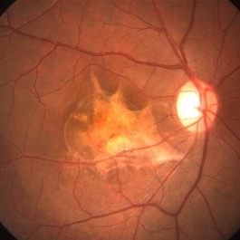
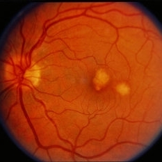
 Myopic Choroidal Neovascular Membrane
Mar 25 2013 by Ratimir Lazic, MD, PhD
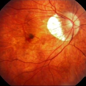
Color fundus photography of a 33-year-old female. In macular area subretinal hemorrhage can be seen. Area of atrophy temporal from PNO. Myopic changes of posterior pole and mid periphery can be noticed. The patient has been treated with 2 consecutive ranibizumab intravitreal injections. BCVA at baseline was 0,05 (Snellen lines) and 0,3 (Snellen lines) 2 months after.
Photographer: Marko Lukic, MD
Imaging device: Zeis Visucam Lite 2
Condition/keywords: high myopia, myopic choroidal neovascularization (CNV), ranibizumab
-
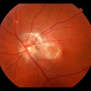
 Punctate Inner Choroidopathy with CNV Treated with Bevacizumab # 6 of 7
Feb 28 2013 by Gregory R. Blaha, MD, PhD
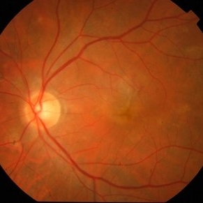
Fundus photo following treatment with bevacizumab in a 31-year-old female with vision loss from a choroidal neovascular membrane (CNV) from punctate inner choroidopathy. The vision improved and was stable following a single injection.
Photographer: Gerard Gauthier, Spindel Eye Assoc., Derry, NH
Imaging device: Zeiss FF 450 Plus
Condition/keywords: bevacizumab, choroidal neovascularization (CNV), punctate inner choroidopathy (PIC)
-
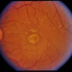
 Punctate Inner Choroidopathy with CNV Treated with Bevacizumab # 1 of 7
Feb 28 2013 by Gregory R. Blaha, MD, PhD
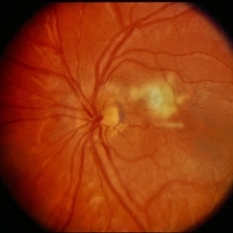
Fundus photograph in a 31-year-old female with vision loss from a choroidal neovascular membrane (CNV) from punctate inner choroidopathy. Note the CNV and hemorrhage superotemporal to the fovea.
Photographer: Gerard Gauthier, Spindel Eye Associates, Derry, NH
Imaging device: Zeiss FF 450 Plus
Condition/keywords: bevacizumab, choroidal neovascularization (CNV), punctate inner choroidopathy (PIC)
-
 Active CNVM
Jul 11 2016 by Manish Nagpal, MD, FRCS (UK), FASRS
Colour photo showing an active CNVM.
Photographer: pooja barot
Condition/keywords: choroidal neovascular membrane (CNVM), optical coherence tomography (OCT)
-
 Astrocytic Hamartoma
Oct 10 2012 by K. Bailey Freund, MD
Fundus photograph of an 85-year-old woman with an astrocytic hamartoma and type 2 choroidal neovascular membrane.
Condition/keywords: choroidal neovascularization (CNV)
-
 Peri-Papillary CNVM
Oct 2 2013 by Jerald A. Bovino, MD
There is a peripapillary cnroidal neovascular membrane visible as the hyperfluorescent area temporal to the disk.
Condition/keywords: peripapillary
-
 Peri-Papillary CNVM
Oct 2 2013 by Jerald A. Bovino, MD
There is a peripapillary cnroidal neovascular membrane visible as the yellow-white areas surrounded by hemorrhage temporal to the disk.
Condition/keywords: peripapillary
-
 Choroidal Osteoma and Secondary Choroidal Neovascular Membrane
Sep 21 2012 by Allen Chiang, MD, FASRS
Fundus photograph of a 44-year old woman with a choroidal osteoma complicated by secondary choroidal neovascular membrane, regressed after serial intravitreal bevacizumab injections. The tumor exhibits areas of decalcification.
Imaging device: Topcon
Condition/keywords: choroidal neovascularization (CNV), choroidal osteoma, macular choroidal osteoma
-
 Punctate Inner Choroidopathy with CNV Treated with Bevacizumab # 2 of 7
Feb 28 2013 by Gregory R. Blaha, MD, PhD
Red-free fundus photograph in a 31-year-old female with vision loss from a choroidal neovascular membrane (CNV) from punctate inner choroidopathy. Note the CNV and hemorrhage superotemporal to the fovea.
Photographer: Gerard Gauthier, Spindel Eye Assoc., Derry, NH
Imaging device: Zeiss FF 450 Plus
Condition/keywords: bevacizumab, choroidal neovascularization (CNV), punctate inner choroidopathy (PIC)
-
---thumb.jpg/image-square;max$300,300.ImageHandler) Punctate Inner Choroidopathy Complicated with CNV
Punctate Inner Choroidopathy Complicated with CNV
Jun 5 2013 by Henry J. Kaplan, MD
A 15-year-old girl presented with blurred vision in left eye (20/200), fundus photography shows multiple deep round hypopigmented scars in the posterior pole with subretinal neovascular membrane #1.
Photographer: Angela Andersson
Condition/keywords: choroidal neovascularization (CNV), punctate inner choroidopathy (PIC)
-
 Peri-Papillary CNVM
Oct 2 2013 by Jerald A. Bovino, MD
There is a peripapillary cnroidal neovascular membrane visible as the hyperfluorescent area temporal to the disk.
Condition/keywords: peripapillary
-
 Punctate Inner Choroidopathy with CNV Treated with Bevacizumab # 4 of 7
Feb 28 2013 by Gregory R. Blaha, MD, PhD
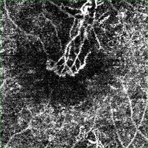
Mid-phase fluorescein angiogram in a 31-year-old female with vision loss from a choroidal neovascular membrane (CNV) from punctate inner choroidopathy.
Photographer: Gerard Gauthier, Spindel Eye Assoc., Derry, NH
Imaging device: Zeiss FF 450 Plus
Condition/keywords: bevacizumab, choroidal neovascularization (CNV), punctate inner choroidopathy (PIC)
-
 Subretinal neovascular membrane
Apr 4 2013 by Jerald A. Bovino, MD
Decolorized blood overlying SRNVM
Condition/keywords: decolorized blood, subretinal neovascular membrane
-
 Active CNVM on Angio OCT
Jul 11 2016 by Manish Nagpal, MD, FRCS (UK), FASRS
Angio OCT picture showing neovascularization corresponding to the area of CNVM.
Photographer: pooja barot
Condition/keywords: choroidal neovascular membrane (CNVM), optical coherence tomography (OCT)
-
 Peripapillary CNVM / Uveitis
Aug 22 2014 by David Callanan, MD
4-year-old patient with peripapillary CNVM / uveitis.
Condition/keywords: choroidal neovascular membrane (CNVM), peripapillary, uveitis
-
 ARMD / Subfoveal CNVM
Feb 13 2015 by David Callanan, MD
Female patient, ARMD / subfoveal CNVM.
Condition/keywords: age-related macular degeneration (AMD), choroidal neovascular membrane (CNVM), subfoveal choroidal neovascularization
-
 Subfoveal Bleed From Extramacular CNVM
Jul 30 2014 by Mallika Goyal, MD
Subfoveal bleed (and fluid on OCT) resolving 4 weeks after intravitreal avastin for the large extrafoveal CNVM that caused the subfoveal bleed with fluid.
Photographer: Mallika Goyal, MD, Apollo Health City, Jubilee Hills, Hyderabad-500033
Condition/keywords: choroidal neovascular membrane (CNVM)
-
 CNVM Secondary to Choroidal Rupture
Jan 26 2017 by Sharat Shivaramaiah Hegde, MS OPHTHALMOLOGY
Young male had history of trauma a year before then came with complaints of dimension of vision in right eye. Vision was 20/200.
Photographer: Anand
Imaging device: zeiss
Condition/keywords: choroidal neovascular membrane (CNVM), choroidal rupture
-
 Subretinal Fibrosis (PPCNVM and POHS) OS
Sep 18 2019 by John S. King, MD
57-year-old white male with history of PPCNVM OS and POHS OU here for a routine visit. History of avastin in 2014, and stable since then. Va OS 20/20. PP scar with macular subretinal fibrosis. No heme or exudates. CR spot supero-nasally.
Photographer: Shelly Blair
Imaging device: Topcon 50
Condition/keywords: choroidal neovascular membrane (CNVM), ocular histoplasmosis syndrome (OHS), peripapillary choroidal neovascularization (PPCNVM), presumed ocular histoplasmosis syndrome (POHS)
-
 ARMD / Subfoveal CNVM
Feb 13 2015 by David Callanan, MD
Female patient, ARMD / subfoveal CNVM.
Condition/keywords: age-related macular degeneration (AMD), choroidal neovascular membrane (CNVM), subfoveal choroidal neovascularization
-
---thumb.jpg/image-square;max$300,300.ImageHandler) Subfoveal Neovascular Membrane And Bridging Fibrotic Band To The Supertemporal Equator
Subfoveal Neovascular Membrane And Bridging Fibrotic Band To The Supertemporal Equator
Oct 4 2013 by Maurice F. Rabb
Acuity OD of 20/800 and OS 20/20. Manifest refraction shows anisometropia: OD +6.50 and OS + 0.75 spheres. The macula of the right eye has a dense fibrotic cicatricial subfoveal scar with mottling of pigment epithelium in the perifoveal area. There is a bridging fibrotic band that extends in the 10 o'clock meridian to the posterior equator tenting up retinal vessels between the two areas. The fluorescein angiogram done has the frames 20-25 reversed on the positive so that they are all of the right eye. The remainder are correctly oriented and show a subfoveal neovascular membrane with surrounding serous fluid overlying residual exudate. There is the fibrotic band that can be seen in the 10:00 o'clock meridian in the late photographs where retinal vessels under traction from the subretinal band leak somewhat.
Condition/keywords: bridging fibrotic band, subfoveal neovascular membrane, supertemporal equator
-
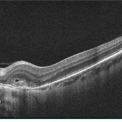 Pseudoxanthoma Elasticum (PXE) with CNV
May 23 2013 by Theodore Leng, MD, MS, FASRS
OCT scan of a 51-year-old woman with pseudoxanthoma elasticum and a leaking choroidal neovascular membrane in the left eye. Fluid is seen on OCT above the CNVM.
Condition/keywords: choroidal neovascularization (CNV), pseudoxanthoma elasticum (PXE)
-
 Punctate Inner Choroidopathy with CNV Treated with Bevacizumab # 3 of 7
Feb 28 2013 by Gregory R. Blaha, MD, PhD
Early-phase fluorescein angiogram in a 31-year-old female with vision loss from a choroidal neovascular membrane (CNV) from punctate inner choroidopathy.
Photographer: Gerard Gauthier, Spindel Eye Assoc., Derry, NH
Imaging device: Zeiss FF 450 Plus
Condition/keywords: bevacizumab, choroidal neovascularization (CNV), punctate inner choroidopathy (PIC)
-
 Presumed Ocular Histoplasmosis Syndrome with Choroidal Neovascular Membrane
Feb 19 2014 by David Callanan, MD
32-year-old woman with presumed ocular histoplasmosis syndrome with choroidal neovascular membrane.
Condition/keywords: presumed ocular histoplasmosis syndrome (POHS)
-
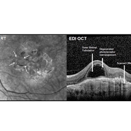 Outer Retinal Tubulation
Mar 27 2018 by Dhaivat Shah
Outer retinal tubulation (ORT) is a feature of photoreceptor rearrangement after chronic retinal damage due to refractory cme, long standing CNVM or old trauma. Photoreceptors lose adhesions to surrounding structures, resulting in outward folding and formation of new lateral contact between photoreceptors to form round structure. They generally remains stable over time. It is important to recognize ORT on OCT because it indicates a refractory state of the pathological condition and poor visual prognosis, and likely not to benefit from any treatment. Here is a case of 62-year-old female with history of 4 previous anti-VEGF injection in left eye for CNVM, with the recent OCT showing formation of ORT with subfoveal scarred membrane.
Photographer: Dr Dhaivat Shah
Condition/keywords: choroidal neovascular membrane (CNVM), outer retinal tubulation
 Loading…