Search results (16 results)
-
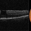
 Macular Pseudohole - OCT
Jan 11 2013 by Gerardo Garcia-Aguirre, MD
OCT scan showing a hyperreflective line that is partially separated from the retina in the fovea and temporal macula, corresponding to an epiretinal membrane. Note the discontinuity of the line just above the fovea, which clinically corresponds to the pseudohole.
Photographer: Gerardo Garcia-Aguirre, MD
Imaging device: Topcon 3DOCT 1000
Condition/keywords: epiretinal membrane (ERM), macular pseudohole
-
 Macular Pseudohole
Jul 7 2015 by Hamid Ahmadieh, MD
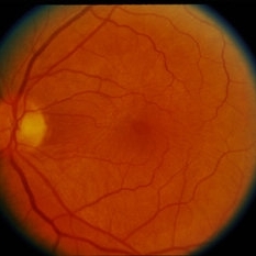
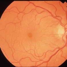
Color fundus photograph and optical coherence tomography of the left eye of a 72-year-old woman with blurred vision due to epiretinal membrane. VA OS is 20/40 . Macular pseudohole is visible.
Photographer: Shabnam Poureh, Negah Eye Center, Tehran, Iran
Imaging device: Topcn
Condition/keywords: color fundus photograph, macular pseudohole, optical coherence tomography (OCT)
-
 OCT of Macular Pseudo Hole
Aug 26 2018 by John T. Thompson, MD
Spectral domain OCT of macular pseudohole with epiretinal membrane.
Imaging device: Heidelberg Spectralis
Condition/keywords: epiretinal membrane (ERM), macular pseudohole, optical coherence tomography (OCT)
-
 Macular Pseudohole
Feb 7 2017 by Manish Nagpal, MD, FRCS (UK), FASRS
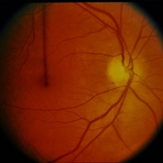
Fundus appearance of a macular hole is basically a small central break in the hyaloid mimicking a macular hole. The OCT in the next picture depicts the break in the hyaloid.
Photographer: Pooja Barot
Condition/keywords: macular pseudohole
-
 Macular Pseudo hole With Schisis
Aug 26 2018 by John T. Thompson, MD
Spectral domain OCT of macular pseudohole with epiretinal membrane and macular schisis. This would be classified as epiretinal membrane and foveoschisis in the international classification system.
Imaging device: Heidelberg Spectralis
Condition/keywords: epiretinal membrane (ERM)
-
 Macular pseudohole - 3D OCT reconstruction
Jan 11 2013 by Gerardo Garcia-Aguirre, MD
3D OCT reconstruction of an epiretinal membrane with a macular pseudohole. Note that the hole is in the epiretinal membrane itself, and not in the retina.
Photographer: Gerardo Garcia-Aguirre, MD
Imaging device: Topcon 3D OCT 1000
Condition/keywords: epiretinal membrane (ERM), macular pseudohole
-
 Macular Pseudo Hole
Aug 26 2018 by John T. Thompson, MD
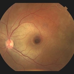
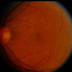
Color fundus photograph of macular pseudohole.
Imaging device: Zeiss FF4
Condition/keywords: macular pseudohole
-
 Macular Pseudohole
Feb 7 2017 by Manish Nagpal, MD, FRCS (UK), FASRS
This OCT reveals the central break in the hyaloid which clinically mimics a macular hole.
Photographer: pooja barot
Condition/keywords: macular pseudohole
-
 ERM with Pseudohole
Oct 1 2014 by David Callanan, MD
66-year-old patient, ERM with pseudohole.
Condition/keywords: epiretinal membrane (ERM), macular pseudohole
-
 ERM with Pseudohole
Oct 1 2014 by David Callanan, MD
66-year-old patient, ERM with pseudohole.
Condition/keywords: epiretinal membrane (ERM), macular pseudohole
-
 Traumatic Pseudohole with Commotio Retinae and Subfoveal Hemorrhage after Blunt Injury
Jun 1 2019 by John S. King, MD
39-year-old African American female with central scotoma two days since blunt head injury in MVA, sent for evaluation of macular hole. 20/150 OS with IOP 12 and no RAPD. No macular hole present. Macular findings include commotio retinae and subfoveal hemorrhage.
Photographer: Stacey Coleman
Imaging device: Topcon
Condition/keywords: blunt trauma, commotio retinae, macular pseudohole, subretinal hemorrhage
-
 ERM with Pseudohole
Oct 1 2014 by David Callanan, MD
66-year-old patient, ERM with pseudohole.
Condition/keywords: epiretinal membrane (ERM), macular pseudohole
-
 Macular Pucker with Pseudohole
Apr 8 2019 by Gary R. Cook, MD, FACS
52-year-old white male with a diffuse ERM and pseudohole OD; V.A. = 20/20-1
Imaging device: Topcon VT-50
Condition/keywords: epiretinal membrane (ERM), idiopathic epiretinal membrane, macular pseudohole, macular pucker
-
 Preretinal Fibrosis
Jan 12 2024 by Virginia Gebhart
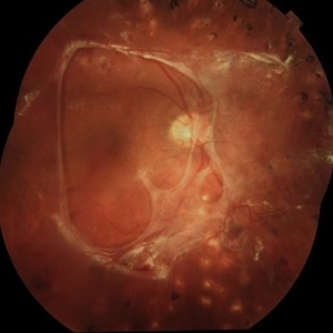
53 year old diabetic male with significant persistent ERM due to fibrotic NV superiorly. Possibly developing a tractional MH. Vitreous Hemorrhage secondary to traction on the fibrosis
Photographer: Virginia Gebhart
Imaging device: Topcon 50DX
Condition/keywords: epiretinal membrane, ERM, fibrosis, macular pseudohole, neovascularization (NV)
-
 Macular Pucker with Pseudohole
Apr 8 2019 by Gary R. Cook, MD, FACS
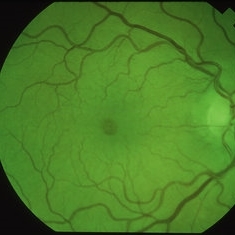
Red-free photograph of a 52-year-old white male with a diffuse ERM and pseudohole OD; V.A. = 20/20-1
Imaging device: Topcon VT-50
Condition/keywords: epiretinal membrane (ERM), idiopathic epiretinal membrane, macular pseudohole, macular pucker
-
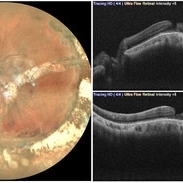 Macular Pseudo-Hole in a Buckled Eye
Sep 10 2021 by Akansha Sharma
Fundus Photograph and Optical Coherence Tomography of left eye showing a macular pseudo-hole in a buckled eye.
Photographer: Dr. Akansha Sharma-Retina Foundation, Ahmedabad
Condition/keywords: macular hole, macular pseudohole, scleral buckle
 Loading…