Search results (73 results)
-
 Chronic Retinal Detachment: Features Slide 1
Oct 22 2012 by Ronald C. Gentile, MD
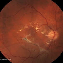
Chronic retinal detachments can be associated with demarcation lines (tidemarks), subretinal bands or sheets, and retinal cysts. Fundus photo of a chronic inferior retinal detachment reveals multiple demarcation lines inferior to the center of the fovea as a result of an inferior temporal dialysis.
Photographer: The New York Eye & Ear Infirmary Department of Medical Imaging
Condition/keywords: chronic retinal detachment, demarcation line
-
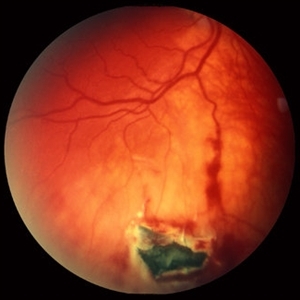 Intraocular Foreign Body, Metallic, in Inferior Retina with Hemorrhage
Oct 1 2012 by Jeffrey G. Gross, MD, FASRS
IOFB, metallic, in inferior retina with hemorrhage.
Condition/keywords: inferior retina, intraocular foreign body
-
 Pars Planitis - Peripheral Uveitis
Nov 9 2012 by Norman Byer
This 25-year-old man had pars planitis, peripheral uveitis bilaterally. In this eye it produced a small tractional oval tear of the retina and an inferior retinal detachment. The typical creamy yellow exudates of pars planitis can be seen in the lower right very close to the ora serrata.
Condition/keywords: creamy yellow exudates, inferior retinal detachment, pars planitis, peripheral uveitis, tractional retinal tear
-
 Chronic Inferior Retinal Detachment
Mar 1 2017 by Philip J. Polkinghorne, MD
Color photograph of chronic retinal detachment with pigment demarcation line and atrophic holes visible. The vision was recorded at 20/20, and follow up is 3 years.
Photographer: Alex Fraser
Condition/keywords: atrophic retinal hole, demarcation line
-
 Toxoplasmosis Slide 2
Oct 22 2012 by Ronald C. Gentile, MD
One month following treatment with Bactrim, Clindamycin, and oral prednisone the focal area chorioretinitis has coalesced with a decrease in overlying vitreous inflammation. Kyrieleis plaques can be seen along the inferior retinal arteriole.
Photographer: The New York Eye & Ear Infirmary Department of Medical Imaging
Condition/keywords: posterior uveitis, toxoplasmosis
-
 Inferior retinal detachment
Dec 19 2012 by Eric A. Postel, MD
Color fundus photograph of an inferior retinal detachment
-
 Intraocular Foreign Body
Apr 9 2014 by Aleksandra V. Rachitskaya, MD, FASRS
Fundus photo of intraocular foreign body located in the inferior retina.
Photographer: Bascom Palmer Eye Institute
Condition/keywords: intraocular foreign body
-
 Inferior RD OD with Exposed Scleral Buckle OD in Previous Images
Feb 4 2013 by James B. Soque, CRA, OCT-C, COA, FOPS
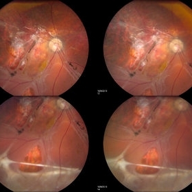
Fundus image of 66-year-old WM with Hx of SBOD in 2009. presents with exposed SBOD and infection seen in accompanying images.
Photographer: James Soque, CRA COA
Imaging device: Topcon TRC 50 DX, MERGE Imaging Software
Condition/keywords: inferior retinal detachment
-
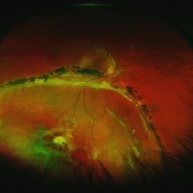
 Coats Disease
Apr 16 2015 by Rita Couceiro, MD, MS
Fundus photograph and fluorescein angiography pictures of a 13-year-old girl with Coats Disease, showing abnormal telangiectatic vessels and intense exsudation in the inferior retinal periphery of the left eye.
Condition/keywords: Coats' disease, retinal telangiectasia
-
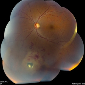
 Rhegmatogenous Retinal Detachment
Oct 11 2013 by Jason S. Calhoun
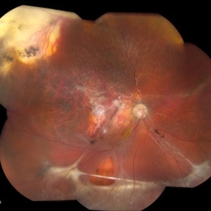
Patient in for a second opinion on RD, right eye. VA is NLP in the right eye. Fundus photography shows inferior retinal detachment with holes and subretinal fibrosis. No further surgery is suggested at this time.
Photographer: Jason S. Calhoun, Ophthalmic Photographer, Department of Ophthalmology, Mayo Clinic Jacksonville
Imaging device: TOPCON TRC 50-EX
-
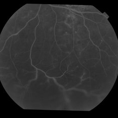
 chronic central serous chorioretinopathy
Oct 31 2012 by Mallika Goyal, MD
Fluorescein angiogram of inferior retina of right eye with chronic CSCR shows dilation of and mild leak from retinal vessels over the inferior serous retinal detachment.
Condition/keywords: central serous chorioretinopathy (CSCR), chronic central serous chorioretinopathy (CSCR), serous retinal detachment
-
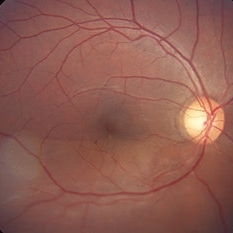
 Sectoral Retinitis Pigmentosa
May 4 2015 by Mallika Goyal, MD
Left fundus of a 46-year-old lady with retinitis pigmentosa affecting inferior retinal quadrants; superior retina, optic nerve head and macula are normal. She is asymptomatic and fundus picture has been stable over 10 years follow-up. She is the offspring of a consanguineous marriage.
Photographer: Mallika Goyal, MD, Apollo Health City, Jubilee Hills, Hyderabad
Condition/keywords: retinal pigmentosa
-
 Rhegmatogenous Retinal Detachment
Oct 11 2013 by Jason S. Calhoun
Patient in for second opinion on RD, right eye. VA is NLP in the right eye. Fundus photography shows inferior retinal detachment with holes and subretinal fibrosis. No further surgery is suggested at this time.
Photographer: Jason S. Calhoun, Ophthalmic Photographer, Department of Ophthalmology, Mayo Clinic Jacksonville
Imaging device: TOPCON TRC 50-EX
-
 Acute Exudative Polymorphous Vitelliform Maculopathy Color OS
Aug 27 2014 by Flavio A. Rezende, MD, PhD
45-year-old man with mild decrease in vision after strong headache. Fundus showing multiple deep irregular vitelliform lesions spread throughout entire posterior pole OU, forming a typical level of subretinal confluent lesions at the inferior retinal vascular arcades. No primary tumor or metastasis found.
Photographer: Eduardo Martins, Pontifícia Universidade Católica - Rio de Janeiro, Brazil
Imaging device: Topcon TRC 50EX
Condition/keywords: polymorphous exudative vitelliform maculopathy
-
 Sectoral Retinitis Pigmentosa
May 4 2015 by Mallika Goyal, MD
Right fundus of a 46-year-old lady with retinitis pigmentosa affecting inferior retinal quadrants; superior retina, optic nerve head and macula are normal. She is asymptomatic and fundus picture has been stable over 10 years follow-up. She is the offspring of a consanguineous marriage.
Photographer: Mallika Goyal, MD, Apollo Health City, Jubilee Hills, Hyderabad
Condition/keywords: retinal pigmentosa
-
 Acute Exudative Polymorphous Vitelliform Maculopathy Color OD
Aug 27 2014 by Flavio A. Rezende, MD, PhD
45-year-old man with mild decrease in vision after strong headache. Fundus showing multiple deep irregular vitelliform lesions spread throughout entire posterior pole OU, forming a typical level of subretinal confluent lesions at the inferior retinal vascular arcades. No primary tumor or metastasis found.
Photographer: Eduardo Martins, Pontifícia Universidade Católica - Rio de Janeiro, Brazil
Imaging device: Topcon TRC 50EX
Condition/keywords: polymorphous exudative vitelliform maculopathy
-
 Sectoral Retinitis Pigmentosa
May 4 2015 by Mallika Goyal, MD
Right fundus of a 46-year-old lady with retinitis pigmentosa affecting inferior retinal quadrants; superior retina, optic nerve head and macula are normal. She is asymptomatic and fundus picture has been stable over 10 years follow-up. She is the offspring of a consanguineous marriage.
Photographer: Mallika Goyal, MD, Apollo Health City, Jubilee Hills, Hyderabad
Condition/keywords: retinal pigmentosa
-
 Sectoral Retinitis Pigmentosa
May 4 2015 by Mallika Goyal, MD
Left fundus of a 46-year-old lady with retinitis pigmentosa affecting inferior retinal quadrants; superior retina, optic nerve head and macula are normal. She is asymptomatic and fundus picture has been stable over 10 years follow-up. She is the offspring of a consanguineous marriage.
Photographer: Mallika Goyal, MD, Apollo Health City, Jubilee Hills, Hyderabad
Condition/keywords: retinal pigmentosa
-
 Choroidal Melanoma through the Pupil
May 28 2016 by Olivia Rainey
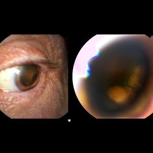
External image of the left eye of a man with metastatic choroidal melanoma, secondary to lung cancer. There was an obstruction of view to the inferior retina, and this prompted the photographer to pull back to see what the problem was.
Photographer: Olivia Rainey
Imaging device: Topcon 50dx
Condition/keywords: choroidal metastasis, color photo, external photography
-
 Retinal Detachment Sparing Fovea By Microns
Sep 24 2018 by samarth mishra
A 29-year-old young female presented with complaint of blurring of vision in the right eye since one year. Best corrected visual acuity was 20/40. On routine examination inferior retinal detachment was noted. Optical coherence tomography (OCT) showed the retinal detachment sparing the fovea by few microns.
Photographer: Aditya Birla Sankara Nethralaya, Kolkata , West Bengal , India
Condition/keywords: color fundus photograph, multicolor, optical coherence tomography (OCT)
-
 Sectoral Retinitis Pigmentosa
May 4 2015 by Mallika Goyal, MD
Left fundus of a 46-year-old lady with retinitis pigmentosa affecting inferior retinal quadrants; superior retina, optic nerve head and macula are normal. She is asymptomatic and fundus picture has been stable over 10 years follow-up. She is the offspring of a consanguineous marriage.
Photographer: Mallika Goyal, MD, Apollo Health City, Jubilee Hills, Hyderabad
Condition/keywords: retinal pigmentosa
-
 chronic central serous chorioretinopathy
Oct 31 2012 by Mallika Goyal, MD
Late phase fluorescein angiogram of inferior retina of left eye with chronic CSCR shows dilation of and mild leak from retinal vessels over the inferior serous retinal detachment.
Photographer: Mallika Goyal, MD
Condition/keywords: central serous chorioretinopathy (CSCR), chronic central serous chorioretinopathy (CSCR)
-
 Retinal Detachment
May 13 2016 by Nichole Lewis
Inferior Retinal Detachment with some demarcation line s/p barrier laser.
Photographer: Nichole Lewis
Condition/keywords: barrier laser
-
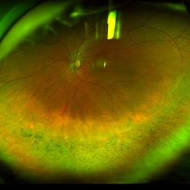 Pigmentary Retinal Dystrophy
May 5 2020 by Olivia Rainey
Ultra-widefield pseudocolor image of an 44-year-old male with pigmentary retinal dystrophy affecting both eyes. He presented with decreased night vision for 6 months prior to his appointment. He stated that his recovery time from transitioning from dark to light areas is reduced. He stated that his peripheral vision has never been very good for most of his life. He admits to environmental hearing loss. Patient denies family history of blin. His vision was 20/20 in both eyes. His full field ERG, visual fields were not consistent with RP. Genetic testing with ID Your IRD and annual follow up has been recommended.
Photographer: Olivia Rainey, OCT-C, COA
Imaging device: Optos California
Condition/keywords: inferior retina, left eye, Optos, pigment, pseudocolor, ultra-wide field imaging
-
 Acute Exudative Polymorphous Vitelliform Maculopathy Red Free OD
Aug 27 2014 by Flavio A. Rezende, MD, PhD
45-year-old man with mild decrease in vision after strong headache. Fundus showing multiple deep irregular vitelliform lesions spread throughout entire posterior pole OU, forming a typical level of subretinal confluent lesions at the inferior retinal vascular arcades. No primary tumor or metastasis found.
Photographer: Eduardo Martins, Pontifícia Universidade Católica - Rio de Janeiro, Brazil
Imaging device: Topcon TRC 50EX
Condition/keywords: polymorphous exudative vitelliform maculopathy
 Loading…