Search results (35 results)
-
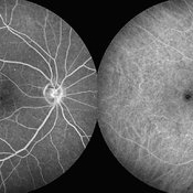
 Type 2 Parafoveal Telangiectasia
Aug 23 2012 by Gerardo Garcia-Aguirre, MD
Fundus photograph showing pigment migration and crystals in the temporal aspect of the fovea.
Photographer: Ricardo Montoya, Asociación para Evitar la Ceguera en México
Imaging device: Zeiss FF4
Condition/keywords: crystals, fovea, idiopathic macular telangiectasia, pigment migration
-
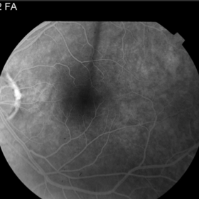
 Macular Telangiectasia Type 2 & CNV
Sep 22 2012 by Hamid Ahmadieh, MD
Color fundus photograph & OCT imagings of the left eye of a 70-year-old man with idiopathic macular telangiectasia type 2 and CNV.
Photographer: Hamid Ahmadieh, MD, Ophthalmic Research Center, Labbafinejad Medical Center, Shahid Beheshti University of Medical Sciences
Imaging device: Topcon Fundus Camera & Topcon OCT
Condition/keywords: choroidal neovascularization (CNV), idiopathic macular telangiectasia, optical coherence tomography (OCT)
-
 Type 2 Parafoveal Telangiectasia
Aug 23 2012 by Gerardo Garcia-Aguirre, MD
Fundus photograph showing pigment migration and crystals in the temporal aspect of the fovea.
Photographer: Noemí Hernández, Asociación para Evitar la Ceguera en México
Imaging device: Zeiss FF4
Condition/keywords: crystals, idiopathic macular telangiectasia, pigment migration
-
 Macular Telangiectasia Type 2
Sep 22 2012 by Hamid Ahmadieh, MD
FA and ICG angiography imagings of the right eye of a 70-year-old man with idiopathic macular telangiectasia type 2.
Photographer: Hamid Ahmadieh, MD, Ophthalmic Research Center, Labbafinejad Medical Center, Shahid Beheshti University of Medical Sciences
Imaging device: HRA
Condition/keywords: idiopathic macular telangiectasia, indocyanine green (ICG) angiography
-
 Macular Telangiectasia Type 2
Sep 22 2012 by Hamid Ahmadieh, MD
Autofluorescence imagings of both eyes of a 70-year-old man with idiopathic macular telangiectasia type 2.
Photographer: Hamid Ahmadieh, MD, Ophthalmic Research Center, Labbafinejad Medical Center, Shahid Beheshti University of Medical Sciences
Imaging device: HRA
Condition/keywords: autofluorescence imaging, idiopathic macular telangiectasia
-
 Macular Telangiectasia Type 2
Sep 22 2012 by Hamid Ahmadieh, MD
Late phase FA and ICG angiography imagings of the right eye of a 70-year-old man with idiopathic macular telangiectasia type 2.
Photographer: Hamid Ahmadieh, MD, Ophthalmic Research Center, Labbafinejad Medical Center, Shahid Beheshti University of Medical Sciences
Imaging device: HRA
Condition/keywords: idiopathic macular telangiectasia, indocyanine green (ICG) angiography
-
 Macular Telangiectasia Type 2 & CNV
Sep 22 2012 by Hamid Ahmadieh, MD
FA and ICG angiography imagings of the left eye of a 70-year-old man with idiopathic macular telangiectasia type 2 and CNV.
Photographer: Hamid Ahmadieh, MD, Ophthalmic Research Center, Labbafinejad Medical Center, Shahid Beheshti University of Medical Sciences
Imaging device: HRA
Condition/keywords: choroidal neovascularization (CNV), idiopathic macular telangiectasia, indocyanine green (ICG) angiography
-
 Macular Telangiectasia Type 2 & CNV
Sep 22 2012 by Hamid Ahmadieh, MD
Late phase FA and ICG angiography imagings of the left eye of a 70-year-old man with idiopathic macular telangiectasia type 2 and CNV.
Photographer: Hamid Ahmadieh, MD, Ophthalmic Research Center, Labbafinejad Medical Center, Shahid Beheshti University of Medical Sciences
Imaging device: HRA
Condition/keywords: choroidal neovascularization (CNV), idiopathic macular telangiectasia, indocyanine green (ICG) angiography
-
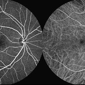
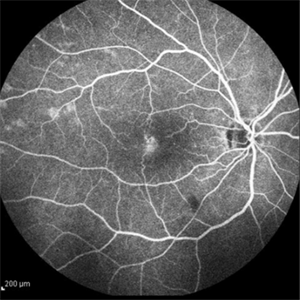
 Telangiectasia
Sep 16 2012 by Ivan R. Batlle, MD
Mid phase fluorescein angiogram of 58-year-old female with decreased vision
Condition/keywords: idiopathic macular telangiectasia
-
 Telagiectasia
Sep 16 2012 by Ivan R. Batlle, MD
Color photograph of 58-year-old female with decreased vision
Condition/keywords: idiopathic macular telangiectasia
-
 Idiopathic Juxtafoveal Telangiectasia, Type 2
Nov 6 2014 by Thomas A. Ciulla, MD, MBA, FASRS
Note the telangiectactic vessels just temporal to the FAZ.
Photographer: Thomas Steele
Condition/keywords: idiopathic macular telangiectasia, juxtafoveal telangiectasis, parafoveal telangiectasia
-
 Idiopathic Juxtafoveal Telangiectasia, Type 2
Nov 6 2014 by Thomas A. Ciulla, MD, MBA, FASRS
Note the telangiectactic vessels just temporal to the FAZ.
Photographer: Thomas Steele
Condition/keywords: idiopathic macular telangiectasia, juxtafoveal telangiectasis, parafoveal telangiectasia
-
 Idiopathic Juxtafoveal Telangiectasia, Type 2
Nov 6 2014 by Thomas A. Ciulla, MD, MBA, FASRS
Note the telangiectactic vessels just temporal to the FAZ.
Photographer: Thomas Steele
Condition/keywords: idiopathic macular telangiectasia, juxtafoveal telangiectasis, parafoveal telangiectasia
-
 Idiopathic Juxtafoveal Telangiectasia, Type 2
Nov 6 2014 by Thomas A. Ciulla, MD, MBA, FASRS
Note the characteristic pseudocyst on OCT.
Photographer: Thomas Steele
Condition/keywords: idiopathic macular telangiectasia, juxtafoveal telangiectasis, parafoveal telangiectasia
-
 Idiopathic Juxtafoveal Telangiectasia, Type 2
Nov 6 2014 by Thomas A. Ciulla, MD, MBA, FASRS
Note the characteristic pseudocyst on OCT.
Photographer: Thomas Steele
Condition/keywords: idiopathic macular telangiectasia, juxtafoveal telangiectasis, parafoveal telangiectasia
-
 Idiopathic Juxtafoveal Telangiectasia, Type 2
Nov 6 2014 by Thomas A. Ciulla, MD, MBA, FASRS
Note the telangiectactic vessels just temporal to the FAZ.
Photographer: Thomas Steele
Condition/keywords: idiopathic macular telangiectasia, juxtafoveal telangiectasis, parafoveal telangiectasia
-
 Idiopathic Juxtafoveal Telangiectasia, Type 2
Nov 6 2014 by Thomas A. Ciulla, MD, MBA, FASRS
Note the telangiectactic vessels just temporal to the FAZ.
Photographer: Thomas Steele
Condition/keywords: idiopathic macular telangiectasia, juxtafoveal telangiectasis, parafoveal telangiectasia
-
 Idiopathic Juxtafoveal Telangiectasia, Type 2
Nov 6 2014 by Thomas A. Ciulla, MD, MBA, FASRS
Note the telangiectactic vessels just temporal to the FAZ.
Photographer: Thomas Steele
Condition/keywords: idiopathic macular telangiectasia, juxtafoveal telangiectasis, parafoveal telangiectasia
-
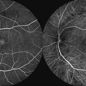
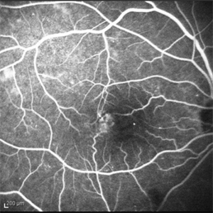
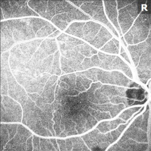
 Idiopathic Juxtafoveal Telangectasia Type 1
Oct 20 2015 by Thomas A. Ciulla, MD, MBA, FASRS
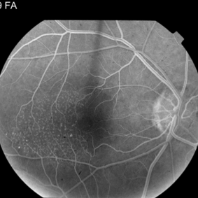
The telangiectasis occurs unilaterally in the temporal half of the macula in an area of 1–2 disc diameters. The anomalies are note in this early frame of the angiogram.
Photographer: Charlotte Harris
Condition/keywords: idiopathic macular telangiectasia, juxtafoveal telangiectasis, parafoveal telangiectasia
-
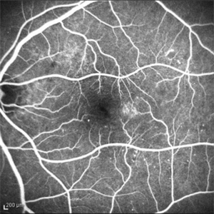
 Idiopathic Juxtafoveal Telangectasia Type 1
Oct 20 2015 by Thomas A. Ciulla, MD, MBA, FASRS
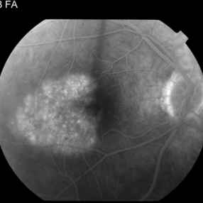
The telangiectasis occurs unilaterally in the temporal half of the macula in an area of 1–2 disc diameters. The late phase of the angiogram shows further leakage temporal to the fovea. Visual loss is mainly caused by macular edema and exudation.
Photographer: Charlotte Harris
Condition/keywords: idiopathic macular telangiectasia, juxtafoveal telangiectasis, parafoveal telangiectasia
-
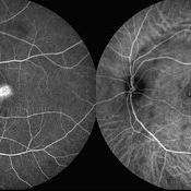
 Coats' Disease Stage 2A
Jun 25 2020 by Thirumalesh Mochi Basavaraj, MD
Fundus photograph (montage) of 9-year-old child with macular exudation. Telangiectic vessels seen. Please note saccular and beaded aneurysmal dilatation of vessels temporally.
Photographer: Puttaswamy
Imaging device: DRI OCT Triton SSOCT- Topcon
Condition/keywords: Coats' disease, idiopathic macular telangiectasia, macular exudates
-
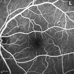
 Idiopathic Juxtafoveal Telangectasia Type 1
Oct 20 2015 by Thomas A. Ciulla, MD, MBA, FASRS
The telangiectasis occurs unilaterally in the temporal half of the macula in an area of 1–2 disc diameters. The anomalies begin to leak in this mid frame of the angiogram.
Photographer: Charlotte Harris
Condition/keywords: idiopathic macular telangiectasia, juxtafoveal telangiectasis, parafoveal telangiectasia
-
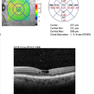
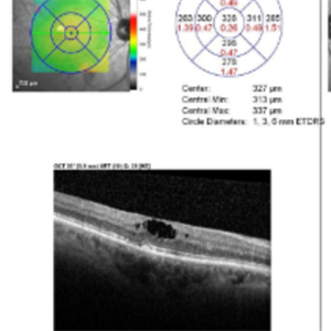
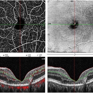 Macular Telangiectasis
May 13 2019 by Hashim Ali Khan, OD, FAAO
OCT-angio of superficial vascular network and structural OCT of a 60-years-old female demonstrating macular TEL showing alterations in FAZ and vascular remodeling and increased the intercapillary distance.
Imaging device: Optical Coherence Tomography Angiography
Condition/keywords: idiopathic macular telangiectasia, macular telangiectasia, macular telangiectasia type 2
-
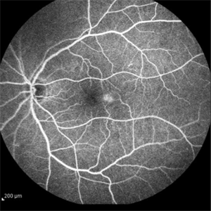
 Idiopathic Juxtafoveal Telangectasia Type 1
Oct 20 2015 by Thomas A. Ciulla, MD, MBA, FASRS
The telangiectasis occurs unilaterally in the temporal half of the macula in an area of 1–2 disc diameters. Vascular anomalies are noted on this red free image.
Photographer: Charlotte Harris
Condition/keywords: idiopathic macular telangiectasia, juxtafoveal telangiectasis, parafoveal telangiectasia
-
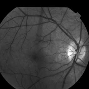
 Idiopathic Juxtafoveal Telangectasia Type 1
Oct 20 2015 by Thomas A. Ciulla, MD, MBA, FASRS
The fellow eye was unremarkable on fluorescein angiography.
Photographer: Charlotte Harris
Condition/keywords: idiopathic macular telangiectasia, juxtafoveal telangiectasis, parafoveal telangiectasia
 Loading…