Search results (76 results)
-
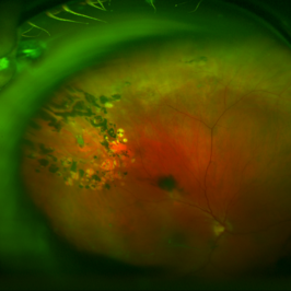
 Horseshoe Tear, In Stereo
Sep 28 2012 by Michael P. Kelly, FOPS
Horse shoe tear, stereo.
Photographer: Michael P. Kelly, FOPS Director, Duke Eye Labs, Duke University Hospital, Duke Eye Center, Durham, NC
Imaging device: Zeiss FF3C
Condition/keywords: stereo pair
-
---thumb.JPG/image-square;max$300,300.ImageHandler) Retinal Detachment with Horseshoe Tear
Retinal Detachment with Horseshoe Tear
Jul 11 2013 by Jason S. Calhoun
Fundus photo shows horseshoe tear superior-temporal in the left eye with retinal detachment.
Photographer: Jason S. Calhoun, Department of Ophthalmology, Mayo Clinic Jacksonville, Florida
-
 Symptomatic Horseshoe Tear
Nov 9 2012 by Norman Byer
This is a fresh, symptomatic horseshoe tear at the site of a cystic retinal tuft in a 63-year-old man. This is really a double horseshoe tear because the two retinal vessels have resisted the vitreal retinal traction and have preserved an intact bridge of tissue between the tears. Note the prominent vitreous condensation attached to the apex of the upper tear and made much more visible because it is seen superimposed over the dark underlying shadow of scleral indentation.
Condition/keywords: bridge of tissue between tears, cystic retinal tuft, scleral indentation, vitreous condensation
-
---thumb.JPG/image-square;max$300,300.ImageHandler) Horseshoe Tear With Laser Treatment
Horseshoe Tear With Laser Treatment
Jul 13 2013 by Jason S. Calhoun
Retinal tear which was treated with a laser retinopexy.
Photographer: Jason S. Calhoun, Department of Ophthalmology, Mayo Clinic Jacksonville, Florida
Condition/keywords: laser retinopexy, retinal tear
-
 Horseshoe Tear
Nov 9 2012 by Norman Byer
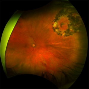
This horseshoe tear was the cause of the detachment in this 54-year-old man. The orange area on the right half of the slide represents the area of scleral indentation. Please note that most of the tear lies over the indented area and appears orange. However, the extreme left side of the tear is brownish black in color because it is exactly superimposed over the dark shadow that always lies just beyond the indented area. The ability of scleral indentation to produce this color change combined with a sharp demarcation between the blackish area and the yellowish edge of intact retina is a pathognomonic sign of a full thickness retinal break.
Condition/keywords: scleral indentation
-
 Sudden Posterior Vitreous Detachment
Nov 9 2012 by Norman Byer
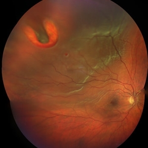
This 52-year-old woman suffered a sudden posterior vitreous detachment which caused a large horseshoe tear at 12:00 o’clock in this eye. It also produced another change at 8:45 in this left eye shown in this photograph. Note the small hemorrhage just to the left of the vessel. Immediately to the left of the hemorrhage and lying alongside of the vessel is a yellowish lesion which actually represents a cystic retinal tuft. You will see it better in the next slide pair.
Condition/keywords: cystic retinal tuft, posterior vitreous detachment, retinal hemorrhage, retinal vessel
-
 Vitreous Hemorrhage
Nov 9 2012 by Norman Byer
This 60-year-old man suddenly developed a vitreous hemorrhage from this acute horseshoe tear 3½ years following cataract extraction when a posterior vitreous detachment occurred. The white nubbin identifies this lesion as a preexisting cystic retinal tuft. The pigment spot beneath the flap is evidence of secondary trophic changes in the pigment epithelium. Note the irregular shape of the flap with the narrow tip and broad base. This was caused by vitreous traction which was exerted at two separate points on the retina and which tore the retina at each place.
Condition/keywords: acute posterior vitreous detachment, irregularly shaped flap, trophic pigmented changes, vitreous hemorrhage, vitreous traction, white retinal tuft
-
 Acute Retinal Detachment
Nov 9 2012 by Norman Byer
This 54-year-old man was referred because of sudden symptoms in his opposite eye in which he had suffered an acute retinal detachment secondary to a horseshoe tear around lattice degeneration. During the examination, the fellow eye shown here was also found to have this large horseshoe tear about 1 o’clock hour (4 disc diameters) in size. A tear occurred around a lattice lesion which is present on the flap but is out of focus. This tear had been asymptomatic even though it was caused by a posterior vitreous detachment and illustrates that even very large tears may produce no symptoms or mild symptoms that are easily overlooked.
Condition/keywords: lattice degeneration, posterior vitreous detachment
-
 Posterior Vitreous Detachment
Nov 9 2012 by Norman Byer
This 68-year-old woman had a recent posterior vitreous detachment which produced this symptomatic horseshoe tear exactly at the site of this cystic retinal tuft. Note the characteristic discrete white nubbin at the apex, which is produced by a cap of glial cells with densely packed cytoplasm.
Condition/keywords: cystic retinal tuft, glial cells, posterior vitreous detachment, white retinal tuft
-
---thumb.JPG/image-square;max$300,300.ImageHandler) Horseshoe Tear Before Laser Treatment
Horseshoe Tear Before Laser Treatment
Jul 13 2013 by Jason S. Calhoun
Retinal tear temporally, proceeded with laser retinopexy.
Photographer: Jason S. Calhoun, Department of Ophthalmology, Mayo Clinic Jacksonville, Florida
Condition/keywords: retinal tear
-
 Horseshoe Tear With Scleral Buckle
Oct 31 2013 by Jason S. Calhoun
Patient had a retinal detachment with retinal tear superior temporally. Underwent surgery and had a scleral buckle placed with good support of the tear. VA is count fingers and will return in 2-months for follow up.
Photographer: Jason S. Calhoun, Ophthalmic Photographer, Department of Ophthalmology, Mayo Clinic Jacksonville
Imaging device: TOPCON TRC 50-EX
Condition/keywords: retinal tear, scleral buckle
-
 Laser Photocoagulation
Nov 9 2012 by Norman Byer
This shows the same lesion 11 days following laser photocoagulation. Still more new hemorrhages can now be seen, and the retinal tissue in the center of the lesion is being visibly pulled forward. If you look carefully, you can see the sharp lower edge of a developing tractional horseshoe tear.
Condition/keywords: laser photocoagulation, retinal tissue, vitreous traction
-
 Retinal Detachment
Nov 9 2012 by Norman Byer
This 50-year-old woman accidentally hit her right eye with a metal tube. The next day she first noticed symptoms of this retinal detachment, which was caused by this usually long and narrow horseshoe tear. Note the rolled edges on each side of the tear and the long blood vessel running down the length of the flap. This illustrates the rule that when traction is exerted on the retina, the retinal blood vessels have a relative resistance to tearing and are the last areas to rupture.
Condition/keywords: retinal vessel, rolled edges of retina
-
 Sub Retinal Gas 1 Day Post Pneumatic Retinopexy
Jul 8 2016 by Asaf Friehmann
Fundus photograph of an 71-year-old male with a sub retinal C3F8 1 day after pneumatic retinopexy for the treatment of rhegmatogenous retinal detachment involving a single 1 hour horseshoe tear at 12 o'clock.
Photographer: Lilach Gorek
Condition/keywords: pneumatic retinopexy
-
 Horseshoe Tear
Oct 12 2017 by Theodore Leng, MD, MS, FASRS
Horseshoe Tear
-
 Horseshoe Tear
Jun 24 2015 by Andree Henaine-Berra, MD
Photograph of the right eye of a 58-year-old male patient with a retinal detachment due to a peripheral horseshoe tear, showing the moment when cryotherapy is applied during the scleral bluckling procedure.
Photographer: Jorge Morales, MD. Hospital General "Dr. Manuel Gea Gonzalez". Mexico City.
Condition/keywords: acute retinal detachment, cryotherapy, scleral buckle
-
 Superior Bullous Detachment With Horseshoe Tear
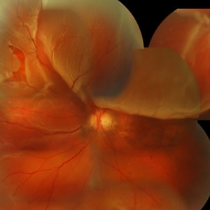
May 15 2014 by Manish Nagpal, MD, FRCS (UK), FASRS
Patient having bullous superior retinal detachment with a horseshoe tear.
Photographer: pooja barot, Optometrist, Retina Foundation, Ahmedabad
-
---thumb.JPG/image-square;max$300,300.ImageHandler) Horseshoe Tear With Detachment
Horseshoe Tear With Detachment
Jul 13 2013 by Jason S. Calhoun
Retinal tear with retinal detachment.
Photographer: Jason S. Calhoun, Department of Ophthalmology, Mayo Clinic Jacksonville, Florida
-
 Horseshoe Tear
Sep 17 2015 by Jason S. Calhoun
Horseshoe tear with sub retinal fluid present superior temporal in the left eye.
Photographer: Jason Calhoun, Mayo Clinic, Department of Ophthalmology
Imaging device: OPTOS 200TX
-
 Rhegmatogenous Retinal Detachment
Jul 14 2016 by PAVEL FLORES-MORENO
Superior horseshoe tear with rhegmatogenous retinal detachment macula off.
Photographer: Pavel Flores-Moreno
Imaging device: Smartphone
-
 Intraoperative Photo Taken During Vitrectomy
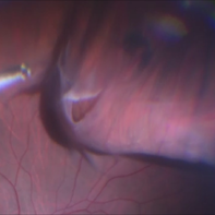
Jan 26 2017 by Manish Nagpal, MD, FRCS (UK), FASRS
Intraoperative photo while doing vitectomy near a horseshoe tear to clear the adherent vitreous enhanced by peripheral scleral indentation while using chandelier light.
Photographer: Manish Nagpal
Imaging device: Still captured from a 3 chip HD camera on microscope
Condition/keywords: cutter, scleral indentation, vitrectomy, vitreous
-
 Rhegmatogenous Retinal Detachment
Mar 3 2021 by Patrik Rajs
A 51-year-old female patient presented with inferior nasal scotoma and 5/10 vision in the right eye due to a retinal detachment with a giant retinal horseshoe tear.
Photographer: Patrik Rajs, EYE CLINIC of Jan Evangelista Purkyne University and Masaryk Hospital, Czech Republic, Ústí nad Labem
Imaging device: Clarus 700
Condition/keywords: giant retinal tear
-
 Retinal Detachment With Macula Detached
Jul 15 2013 by Jason S. Calhoun
Patient went to the ER over the weekend with loss of vision in the right eye. VA was count fingers in the right eye. Noticed a curtain in the right eye. Horseshoe tear was seen at 10-o'clock temporally, right eye. Proceeded with surgery the next day.
Photographer: Jason S. Calhoun, Department of Ophthalmology, Mayo Clinic Jacksonville, Florida
Imaging device: TOPCON TRC 50-EX
-
 Horseshoe Tear
Sep 18 2017 by Theodore Leng, MD, MS, FASRS
Horseshoe tear treated with laser
Condition/keywords: laser, laser retinopexy
 Loading…