Search results (131 results)
-
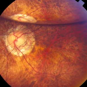
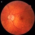
 Inferior Rhegmatogenous Retinal Detachment with Subretinal Fibrosis
Aug 23 2012 by Gabriela Lopezcarasa Hernandez, MD
Asymptomatic 25-year-old woman with high myopia.
Photographer: Gabriela Lopezcarasa Hernandez, Hospital Angeles Lomas
Imaging device: FF4
Condition/keywords: high myopia, subretinal fibrosis
-
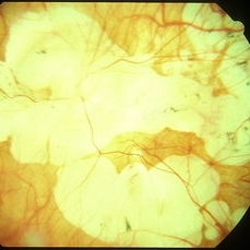
 "Internal Mirroring" Effect by Intraocular Gas
Mar 25 2014 by Homayoun Tabandeh, MD, FASRS
"Internal mirroring" by residual intraocular gas in a highly myopic patient 3 weeks post repair of retinal detachment with pars plana vitrectomy and C3F8 gas.
Photographer: Danny Rivas
Condition/keywords: high myopia, intraocular gas
-
 Myopic CNV
Jan 11 2013 by Alex P. Hunyor, MD
Myopic macular degeneration complicated by subretinal neovascularisation, left eye.
Condition/keywords: high myopia, myopia, myopic choroidal neovascularization (CNV)
-
 Myopic Choroidal Neovascular Membrane
Mar 25 2013 by Ratimir Lazic, MD, PhD
Color fundus photography of a 33-year-old female. In macular area subretinal hemorrhage can be seen. Area of atrophy temporal from PNO. Myopic changes of posterior pole and mid periphery can be noticed. The patient has been treated with 2 consecutive ranibizumab intravitreal injections. BCVA at baseline was 0,05 (Snellen lines) and 0,3 (Snellen lines) 2 months after.
Photographer: Marko Lukic, MD
Imaging device: Zeis Visucam Lite 2
Condition/keywords: high myopia, myopic choroidal neovascularization (CNV), ranibizumab
-
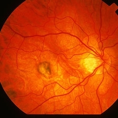
 Peripapillary Atrophy With High Myopia
Feb 4 2015 by H. Michael Lambert, MD
Peripapillary atrophy and central macular degeneration seen in high myopia.
Condition/keywords: high myopia, peripapillary atrophy
-
 Fuch's Spot
Apr 2 2019 by Gary R. Cook, MD, FACS
20-year-old patient with high myopia and a Fuch's spot OD.
Condition/keywords: Fuchs, high myopia, pathologic myopia
-
 Giant Retinal Tear
Oct 9 2012 by Audina M. Berrocal, MD FASRS
Teenager with high myopia and a GRT
Photographer: Ditte Hess CRA, BPEI
Imaging device: Fundus Camera
Condition/keywords: high myopia, retinal degeneration, retinal tear
-
 Peripapillary Atrophy With High Myopia
Feb 4 2015 by H. Michael Lambert, MD
Peripapillary atrophy and central macular degeneration seen in high myopia.
Condition/keywords: high myopia, peripapillary atrophy
-
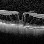 Myopic Macular Schisis with Lamellar Macular Hole
May 26 2014 by John T. Thompson, MD
Spectral domain OCT of patient with high myopia and myopic macular schisis resulting in lamellar macular hole.
Condition/keywords: lamellar macular hole, myopic macular schisis
-
 Myopic Shift With Tilted Optic Disc.
Jul 11 2013 by Jason S. Calhoun
Black female who has a myopic shift shows tilted optic disc in the right eye.
Photographer: Jason S. Calhoun, Department of Ophthalmology, Mayo Clinic Jacksonville, Florida
Condition/keywords: high myopia
-
 Myopic CNV
May 2 2013 by Henry J. Kaplan, MD
Subretinal membrane in high myopia.
Condition/keywords: myopic choroidal neovascularization (CNV)
-
 Progressive Bifocal Chorioretinal Atrophy
Feb 1 2015 by Andree Henaine-Berra, MD
Fundus photograph of the left eye of an 13-year-old female patient with poor vision, high myopia and nystagmus. The image shows macular dragging, a limited area of chorioretinal atrophy temporal to the optic disc and an extense area of chorioretinal atrophy temporal to the macula that extended to the extreme periphery.
Photographer: Andree Henaine-Berra, MD
Condition/keywords: chorioretinal atrophy
-
 Dome-Shaped Macula With Subretinal Fluid
Jun 14 2018 by Gerardo Garcia-Aguirre, MD
EDI OCT of the right eye of a 17-year-old highly myopic girl. Subfoveal fluid is present. There is choroidal thinning, and scleral thickening in the foveal area.
Photographer: Gerardo Garcia-Aguirre, MD
Imaging device: Heidelberg Spectralis
Condition/keywords: dome shaped macula, high myopia
-
 High Myopia
May 2 2013 by Henry J. Kaplan, MD
Chorioretinal atrophy in high myopia and tilted disc.
Condition/keywords: high myopia, tilted disc
-
 White Without Pressure
Mar 13 2020 by Anfisa Ayalon, MD
Fundus photograph of a 26-year-old woman with high myopia. Note inferotemporally margins of sharply demarcated WWP area.
Photographer: Anfisa Ayalon, MD., Meir Medical Center, Kfar Saba, Israel.
Imaging device: California, Optos 200 DTX
Condition/keywords: myopia, white without pressure
-
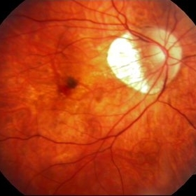
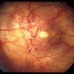
 Spontaneous Macular Hemorrhage in High Myopia
Jul 7 2015 by Hamid Ahmadieh, MD
A highly myopic 30-year-old-man noticed sudden drop of vision and metamorphopsia in his left eye. Color fundus photograph showed a macular haemorrhage (Fig. a) which partially resolved spontaneously after 6 weeks ( Fig. b). Four months later, VA improved to 20/40 and the image distortion markedly resolved ( Fig. c). Lacquer cracks were visible.
Photographer: Soulmaz Shahmohammad, Negah Eye Center, Tehran, Iran
Condition/keywords: color fundus photograph, high myopia, macular hemorrhage
-
---thumb.JPG/image-square;max$300,300.ImageHandler) High Myopia with CNVM
High Myopia with CNVM
Dec 1 2013 by Mallika Goyal, MD
22-year-old male with bilateral high myopia with macular degeneration. Left eye has CNVM with bleed.
Photographer: Mallika Goyal, MD, Apollo Hospitals, Hyderabad, India
Condition/keywords: high myopia
-
 Foveoschisis secondary to high myopia
Mar 13 2015 by Niloofar Piri, MD
Infrared and HD-OCT of the right eye in a 55-year-old African American female with high myopia (more than -6.00 D), BCVA: 20/25 OU Cartwheel appearance of the fovea in the infrared imaging is visible. HD- OCT demonstartes schisis in different layers of the retina (both NFL and OPL; notice stretching of the Muller cells); VMT is also present . Outer retinal layers are preserved which explains the good vision . She had the same findings in OS.
Photographer: Niloofar Piri, MD
Imaging device: Heidelberg Spectralis
Condition/keywords: high myopia, retinoschisis
-
 High Myopia with Lacquer Cracks
Apr 2 2019 by Gary R. Cook, MD, FACS
High myopia with lacquer cracks; -12.25D
Condition/keywords: high myopia, lacquer cracks, myopia
-
---thumb.jpg/image-square;max$300,300.ImageHandler) Congenital Cataracts
Congenital Cataracts
Dec 26 2013 by David Callanan, MD
42-year-old female patient, 20/30 OU; high myope.
Condition/keywords: congenital cataract, high myopia
-
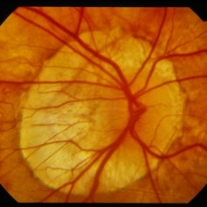
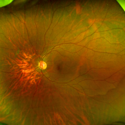
 Posterior Staphylamas
May 2 2013 by Henry J. Kaplan, MD
Highly myopic changes with posterior staphyloma and visible sclera.
Condition/keywords: high myopia, posterior staphyloma
-
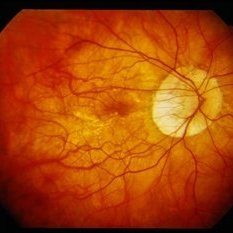
 High Myopia
Jun 14 2018 by Mitzy E Torres Soriano, MD
Fundus photograph (left eye) of a female patient with high myopia, chorioretinal atrophy, pigmentary changes and posterior staphyloma.
Photographer: Mitzy Torres Soriano
Condition/keywords: chorioretinal atrophy, high myopia, posterior staphyloma
-
---thumb.JPG/image-square;max$300,300.ImageHandler) High Myopia with CNVM
High Myopia with CNVM
Dec 1 2013 by Mallika Goyal, MD
CNVM regressing and bleed resolved following anti-VEGF therapy.
Photographer: Mallika Goyal, MD, Apollo Hospitals, Hyderabad, India
Condition/keywords: high myopia
-
 High Myopia
Apr 2 2019 by Gary R. Cook, MD, FACS
17-year-old Vietnamese male with -23D myopia; V.A.= 20/40
Imaging device: Topcon VT-50
Condition/keywords: high myopia, lacquer cracks, pathologic myopia
-
 Myopic Traction Maculopathy
May 31 2014 by Rameez N Hussain, MD
Spectral domain optical coherence tomography of macular detachment in posterior staphyloma - myopic traction maculopathy (MTM).
Photographer: Rameez N Hussain MD, Vitreo Retinal Services, Giridhar Eye Institute, Cochin, India
Imaging device: Heidelberg Spectralis
Condition/keywords: high myopia, macular detachment, myopic traction maculopathy, pathologic myopia, posterior staphyloma
 Loading…