Search results (50 results)
-
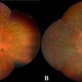
 Gyrate Atrophy of Choroid and Retina
Apr 19 2014 by Mallika Goyal, MD
Right eye fundus of a 45-year-old male patient with advanced gyrate atrophy of the choroid and retina with macular sparing. Optic nerve head is healthy.
Photographer: Mallika Goyal, MD, Apollo Health City, Hyderabad, India
Condition/keywords: choroid, gyrate atrophy
-
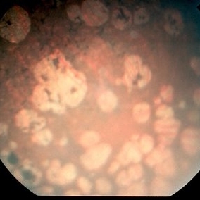
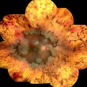
 Gyrate Atrophy
Jan 6 2019 by Hashim Ali Khan, OD, FAAO
Montage of Multiple Fundus Photographs from the right eye of a 25-year-old woman with gyrate atrophy.
Photographer: Ahmed Abbass
Imaging device: Topcon TRC-NW8F
Condition/keywords: gyrate atrophy, hereditary retinal dystrophy, retinal dystrophy
-
 Gyrate Atrophy
Oct 9 2012 by Alan D. Letson, MD
Focal atrophic choroidal lersions in a 12-year-old boy with markedly elevated serum ornithine levels.
Photographer: Beverly Radcliffe
Condition/keywords: gyrate atrophy, ornithine
-
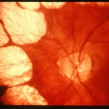
 Gyrate Atrophy
Jun 4 2014 by Neha Goel, MS DNB FRCS (Glasg)
Fundus photograph of the right eye of a 25-year-old female.
Photographer: Neha Goel
Imaging device: Zeiss Visucam
Condition/keywords: gyrate atrophy, hereditary choroidal dystrophy
-
 Gyrate Atrophy
Oct 19 2012 by Larry Halperin, MD
Gyrate atrophy
-
---thumb.jpg/image-square;max$300,300.ImageHandler) Gyrate Atrophy
Gyrate Atrophy
-
---thumb.jpg/image-square;max$300,300.ImageHandler) Gyrate Atrophy
Gyrate Atrophy
Aug 1 2013 by From the Collections of Thomas M. Aaberg, MD and Thomas M. Aaberg Jr., MD
Gyrate atrophy.
Condition/keywords: gyrate atrophy
-
---thumb.jpg/image-square;max$300,300.ImageHandler) Gyrate Atrophy
Gyrate Atrophy
-
---thumb.jpg/image-square;max$300,300.ImageHandler) Gyrate Atrophy
Gyrate Atrophy
-
---thumb.jpg/image-square;max$300,300.ImageHandler) Gyrate Atrophy
Gyrate Atrophy
-
 Gyrate Atrophy
Oct 31 2018 by Dhaivat Shah
50-year-old male came in with complaint of daytime vision loss for a year and nighttime vision loss for more than 20 years, gradually increasing day by day. Fundus showed paving-stone like areas of atrophy of the RPE involving the macula which coalesces to form a characteristic scalloped border at the junction of normal and abnormal RPE. Gyrate atrophy is an autosomal recessive dystrophy caused by tenfold elevations of plasma ornithine, which is toxic to the RPE and choroid. Patients with gyrate atrophy have hyperpigmented fundi, with lobular loss of the RPE and choroid, normally sparing the fovea. The finding of generalized hyperpigmentation of the remaining RPE helps to clinically distinguish gyrate atrophy from choroideremia. Affected patients usually develop night blindness during the first decade of life and experience progressive loss of visual field and visual acuity later in the disease course. Early diagnosis is crucial because treatment in form of Arginine free diet and oral pyridoxine helps in slowing the progression of disease.
Imaging device: Optos
Condition/keywords: fundus autofluorescence (FAF), gyrate atrophy
-
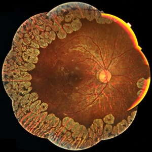
 Gyrate Atrophy of Choroid and Retina
Apr 19 2014 by Mallika Goyal, MD
Right eye fundus of a 45-year-old male patient with advanced gyrate atrophy of the choroid and retina with macular sparing. Optic nerve head is healthy.
Photographer: Mallika Goyal, MD, Apollo Health City, Hyderabad, India
Condition/keywords: choroid, gyrate atrophy
-
---thumb.jpg/image-square;max$300,300.ImageHandler) Gyrate Atrophy
Gyrate Atrophy
-
 Gyrate Atrophy of Choroid and Retina
Apr 19 2014 by Mallika Goyal, MD
Left eye fundus of a 45-year-old male patient with advanced gyrate atrophy of the choroid and retina with macular sparing. Optic nerve head is healthy.
Photographer: Mallika Goyal, MD, Apollo Health City, Hyderabad, India
Condition/keywords: gyrate atrophy
-
 Gyrate Atrophy of Choroid and Retina
Apr 19 2014 by Mallika Goyal, MD
Left eye fundus of a 45-year-old male patient with advanced gyrate atrophy of the choroid and retina with macular sparing. Optic nerve head is healthy.
Photographer: Mallika Goyal, MD, Apollo Hospitals, Hyderabad, India
Condition/keywords: choroid, gyrate atrophy
-
---thumb.jpg/image-square;max$300,300.ImageHandler) Gyrate Atrophy
Gyrate Atrophy
-
---thumb.jpg/image-square;max$300,300.ImageHandler) Gyrate Atrophy
Gyrate Atrophy
-
 Gyrate Atrophy
Sep 23 2020 by Hashim Ali Khan, OD, FAAO
Widefield color fundus image of a young male with gyrate atrophy.
Imaging device: Optomap
Condition/keywords: gyrate atrophy
-
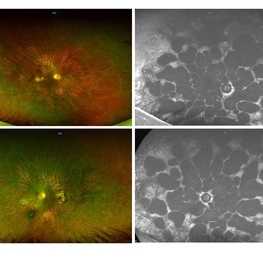
 Gyrate Atrophy
Oct 30 2020 by JEFFERSON R SOUSA, Tecg.º (Biomedical Systems Technology)
Female patient, 28-year-old, with low vision in both eyes since childhood. In routine examination, important changes were observed with atrophic, symmetrical and bilateral aspects with apparently preservation of the central retina.
Condition/keywords: gyrate atrophy
-
 Gyrate
Dec 22 2014 by Howard Schatz, MD
63-year-old white male. Gyrate. 20/30 and 20/30.
Condition/keywords: gyrate atrophy
-
 Gyrate Atrophy of the Choroid and Retina
May 1 2019 by Anmol Naik
A 34-year-old Indian male presented with gradual progressive bilateral diminution of peripheral vision since 6 years. His best corrected visual acuity was 6/60, N36 in right eye and 6/9, N6 in left. Wide-field fundus imaging demonstrated scalloped areas of chorioretinal atrophy with well-defined margins. His plasma ornithine levels were elevated.at 203.9 nmol/ml. Based on the typical features, a diagnosis of gyrate atrophy was made.
Photographer: Anmol Naik, Sankara Nethralaya, Chennai, India
Imaging device: Zeiss CLARUS 500
Condition/keywords: chorioretinal atrophy, gyrate atrophy
-
 Gyrate Atrophy
Apr 12 2023 by Ahmed Abbas Hashmi, OD
Left eye fundus of a 53-year-old male patient with advanced gyrate atrophy of the choroid and retina with macular sparing. Optic nerve head is healthy.
Photographer: Ahmed Abbas Hashmi
Imaging device: Topcon TRC-NW8F
Condition/keywords: chorioretinal atrophy
-
 Gyrate
Dec 22 2014 by Howard Schatz, MD
63-year-old white male. Gyrate. 20/300.
Condition/keywords: gyrate atrophy
 Loading…