Search results (179 results)
-
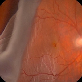
 Giant Retinal Tear
Oct 9 2012 by Audina M. Berrocal, MD FASRS
Teenager with high myopia and a GRT
Photographer: Ditte Hess CRA, BPEI
Imaging device: Fundus Camera
Condition/keywords: high myopia, retinal degeneration, retinal tear
-
 Retinal Tack
Oct 11 2012 by Michael P. Kelly, FOPS
This is a retinal fundus photograph I took in 1987 while working with Howard Schatz, MD and H. Richard McDonald, MD, when retinal tacks were used to repair giant retinal tears. I purposely underexposed the retina because the retinal tack is so highly relective.
Photographer: Michael P. Kelly, FOPS Director, Duke Eye Center Labs, Duke University Hospital
Condition/keywords: retinal tacks, retinal tear
-
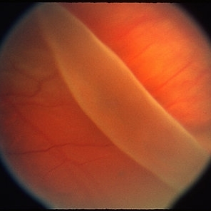
 Giant Retinal Tear Slide 1
Oct 22 2012 by Ronald C. Gentile, MD
Acute loss of vision in a myopic man with flashes and floaters in the right eye. The giant retinal tear is flapped over with the macula detached. The undersurface of the retina can be seen temporally.
Photographer: The New York Eye & Ear Infirmary Department of Medical Imaging
Condition/keywords: retinal tear, vitrectomy
-
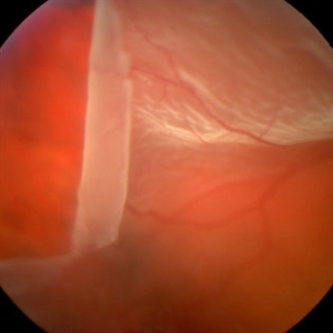 Giant Retinal Tear
Mar 1 2014 by Homayoun Tabandeh, MD, FASRS
Giant retinal tear with radial element and rolled back posterior edge.
Condition/keywords: retinal tear
-
 Giant Retinal Tear Slide 3
Oct 22 2012 by Ronald C. Gentile, MD
Following repair of the retinal detachment with vitrectomy and scleral buckle the edge of the giant retinal tear can be seen to be lying flat on the indentation of the scleral buckle.
Photographer: The New York Eye & Ear Infirmary Department of Medical Imaging
Condition/keywords: retinal tear, vitrectomy
-
---thumb.JPG/image-square;max$300,300.ImageHandler) Giant retinal tear
Giant retinal tear
Oct 26 2012 by Mallika Goyal, MD
Fundus photograph of a 43-year-old gentleman with giant retinal tear and retinal detachment.
Condition/keywords: retinal tear
-
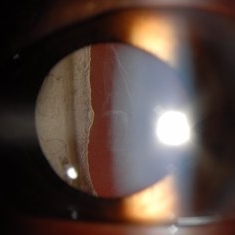 Stickler Syndrome
Sep 28 2016 by Philip J. Polkinghorne, MD
Slit lamp photograph
Photographer: Alex Fraser
Condition/keywords: giant retinal tear, membranous vitreous, Stickler Syndrome
-
 Giant Retinal Tear Slide 2
Oct 22 2012 by Ronald C. Gentile, MD
Following repair of the retinal detachment with vitrectomy and scleral buckle the retina is attached and the patient's vision improved.
Photographer: The New York Eye & Ear Infirmary Department of Medical Imaging
Condition/keywords: retinal tear, vitrectomy
-
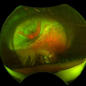
---thumb.jpg/image-square;max$300,300.ImageHandler) Retinectomy With Diathermy in a Giant Tear
Retinectomy With Diathermy in a Giant Tear
Mar 13 2014 by Marcelo Zas, MD PhD
The image show a giant tear in a myopic patient. We use diathermy to avoid intraop bleeding.
Photographer: Marcelo Zas MD PhD
Condition/keywords: giant retinal tear, myopia
-
 "Hang in There"
Apr 20 2021 by Tomas Minelli, MD
Fundus wide field photograph of a 50-year-old man with a macular detachment associated with a big temporal superior tear. The laser is firmly holding the progression of the tear in the 14th day post- laser. BCVA 20/20
Photographer: Livia Conci, Universtity of São Paulo
Imaging device: Optos Daytona
Condition/keywords: giant retinal tear
-
 Giant Retinal Tear
Mar 29 2014 by Min Kim, MD, PhD, MBA, FASRS
Wide field fundus photograph of a 25 year-old male shows giant retinal tear with inverted retinal flap.
Photographer: Young Duk Bae, Yonsei University, Gangnam Severance Hospital
Imaging device: Optomap
Condition/keywords: giant retinal tear
-
 Silicone Oil
Jan 3 2013 by Wilfredo C. Lara, MD
Status post giant retinal tear repair with use of silicone oil.
Condition/keywords: silicone oil
-
 Giant Retinal Tear
May 15 2014 by Manish Nagpal, MD, FRCS (UK), FASRS
Patient presenting with a acute loss of vision with a giant retinal tear.
Photographer: pooja barot, Optometrist, Retina Foundation, Ahmedabad
Condition/keywords: giant retinal tear
-
 Traumatic Retinal Tear
Sep 10 2014 by Mehul A Shah
A myopic male patient 30-years-old presented to outdoor and found to have retinal detachment with giant tear following blunt trauma
Photographer: Drashti Netralaya,Dahod
Imaging device: FF 450
Condition/keywords: giant retinal tear
-
 24 Hours Post Scleral Wound Closure+ Scleral Buckle+25 g Vitrectomy+Silicon Oil
Jan 23 2015 by Carlos Quezada-Ruiz, MD, FASRS
24 hours post op fundus photograph of a 43-year-old man who had perforating injury to the right eye with a small piece of plastic while he was hammering. OD LP, subconjunctival hemorrhage, clear cornea, hyphema, irido and ciclodyalisis as well as a luxated lens with traumatic cataract and a dense vitreous hemorrhage. B-US showed rhegmatogenous retinal detachment with a tear and a big inferior hemorrhagic choroidal detachment. 360 peritomy revealed 2-entry scleral wounds were found in zone II (M V and M VI) and closure was performed. 25 G PPV was performed with the infusion canal placed in the AC through the limbus. Lensectomy and removal of a dense recent vitreous hemorrhage revealed a white detached retina with an exit wound through the temporal inferior segment of the optic nerve with a nasal GRT and sub retinal hemorrhage as well as temporal inferior choroidal, PVD was induced and PFOs helped stabilizing the retina while vitrectomy and sub-retinal hemorrhage was removed through the GRT. Fluid air exchange was made and 360 endolaser over the buckle indentation was done and silicon oil was used as endotamponade. This picture was taken 24 hrs after the surgery.
Photographer: Lilibeth Rodriguez, Instituto de la Visión. Torreon, Mexico.
Condition/keywords: central retinal artery occlusion (CRAO), giant retinal tear, trauma
-
 Giant Retinal Tear
Apr 1 2016 by Nichole Lewis
Giant retinal tear montaged on Anterior Segment due to the Detachment being very bullous.
Photographer: Nichole Lewis - Pennsylvania Retina Specialists, Camp Hill, PA
Condition/keywords: giant retinal tear, retinal tear
-
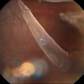
 Giant retinal tear
Oct 26 2012 by Mallika Goyal, MD
Bare choroid in an eye with giant retinal tear.
Condition/keywords: retinal tear
-
---thumb.JPG/image-square;max$300,300.ImageHandler) Giant Retinal Tear Treated With Laser
Giant Retinal Tear Treated With Laser
Jul 8 2013 by Jason S. Calhoun
Patient in with a shower of floaters. VA was 20/30 and fundus exam shows giant retinal tear temporally. Patient was treated with laser retinopexy to prevent retinal detachment.
Photographer: Jason S. Calhoun, Department of Ophthalmology, Mayo Clinic Jacksonville, Florida
Condition/keywords: laser retinopexy, retinal tear
-
 Retinal Detachment with Giant Retinal Tear and Macular Hole
Jan 6 2020 by MATTEO FORLINI, MD
A 61-year-old-male patient presented with sudden diminution of vision in the right eye due to retinal detachment with giant retinal tear and macular hole. Best corrected visual acuity (BCVA) at presentation was 20/200. A 23 G vitrectomy was performed. The edges of the tear were unrolled and complete retinal re-attachment under PFCL was achieved. A 360 degree intraoperative endolaser was performed on the peripheral retina as well as around the edges of the tears. PFCL was exchanged with silicone oil 5000cs as final tamponade. At six-months follow-up retina was attached and macular hole was repaired. Best-corrected visual acuity is 20/125 at present.
Photographer: Matteo Forlini MD, San Marino Hospital, Republic of San Marino
Condition/keywords: full thickness macular hole, giant retinal tear, silicone oil
-
 Giant Retinal Tear
-
---thumb.JPG/image-square;max$300,300.ImageHandler) Giant Retinal Tear
Giant Retinal Tear
Jul 13 2013 by Jason S. Calhoun
Giant retinal tear with retinal detachment.
Photographer: Jason S. Calhoun, Department of Ophthalmology, Mayo Clinic Jacksonville, Florida
Condition/keywords: retinal tear
-
 Giant Retinal Tear With RD
Jun 29 2013 by Jason S. Calhoun
Middle aged patient comes in with sudden loss of vision inferior, nasally. Patient presents plus 2 APD in the right eye. Fundus exam reveals a giant retinal tear at 10-11 o'clock with RD. Vitrectomy with laser and gas exchange of C3F8 scheduled.
Photographer: Jason S. Calhoun, Mayo Clinic Jacksonville, Florida
Imaging device: TOPCON TRC 50-EX
Condition/keywords: retinal tear
-
 Retinal Detachment with Giant Retinal Tear
Mar 9 2013 by Young-Gyun Kim, MD
Fundus photograph of a 45-year-old man with retinal detachment and giant retinal tear.
Photographer: Shin Ji-Young, Eulji university, Seoul
Imaging device: Topcon TRC 50 EX
Condition/keywords: retinal tear
-
 Multiple Retinal Lesions Secondary to Blunt Trauma
Jun 19 2018 by Somnath Chakraborty, MD
A montage of the right eye of a 15-year-old boy, who was struck by a football. The image shows multiple choroidal ruptures in the macular area, with sub-retinal blood and multiple, large retinal tears temporally. There is also an area of juxtapapillary, pigmentary changes.
Photographer: Saptarshi Mehta, Retina Institute of Bengal
Condition/keywords: blunt trauma, choroidal rupture, giant retinal tear, subretinal hemorrhage
-
 Macula-Sparing GRT RRD
Jul 6 2017 by Andrew A. Moshfeghi, MD, MBA, FASRS
Wide-field fundus photograph of a 43-year-old myopic man with a history of lattice retinal degeneration status posterior barrier laser performed elsewhere who presented with a giant-retinal tear associated retinal detachment of the right eye.
Photographer: Jay Jiang, University of Southern California Roski Eye Institute
Imaging device: Optos California
Condition/keywords: acute retinal detachment, giant retinal tear, lattice degeneration
 Loading…