Search results (121 results)
-
 Diabetic Macular Edema, Proliferative Diabetic Retinopathy, Neovascularization Elsewhere, DME, PDR, NVE
Apr 1 2013 by James B. Soque, CRA, OCT-C, COA, FOPS
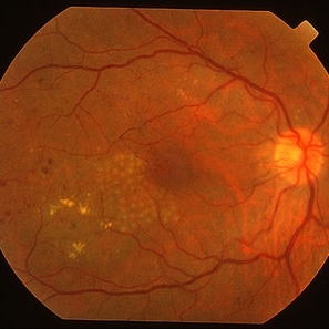
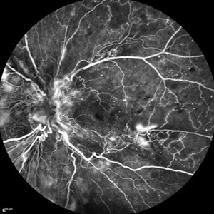
39-year-old white female and long standing diabetis, c/o new peripheral symptoms of left eye. FA OS reveals diabetic macular edema, microaneurysms, and neovasculaization elsewhere. Fluorescein Angogram, Early Phase, 50 Deg, 2x Mag.
Photographer: James B Soque, CRA, COA
Imaging device: Topcon TRC 50DX with MERGE software, OIS 10.6.45
Condition/keywords: diabetic macular edema, neovascularization (NV), proliferative diabetic retinopathy (PDR)
-
 Diabetic Retinopathy Hard Exudates OS
Jun 30 2013 by Rogerio N Shinsato, MD, PhD
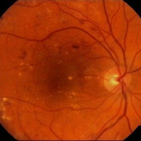
Fundus photograph with diabetic retinopathy.
Condition/keywords: diabetic macular edema, foveal hard exudates
-
 MPC for CSME
Mar 29 2013 by Henry J. Kaplan, MD
Right after MPC for CSME in diabetes (before the introduction of anti-VEGFs).
Condition/keywords: clinically significant macular edema (CSME), diabetic macular edema, multifocal chorioretinitis (MCP)
-
---thumb.jpg/image-square;max$300,300.ImageHandler) Diabetic Retinopathy Hard Exudates OD
Diabetic Retinopathy Hard Exudates OD
Jun 30 2013 by Rogerio N Shinsato, MD, PhD
Fundus photograph with diabetic retinopathy.
Condition/keywords: diabetic macular edema, foveal hard exudates
-
 Diabetic Macular Edema
Oct 12 2012 by Gregg T. Kokame, MD, MMM, FASRS
Diabetic macular edema
Photographer: Jaclyn Pisano, Retina Consultants of Hawaii
Imaging device: Zeiss FF-450 plus
Condition/keywords: diabetic macular edema
-
---thumb.JPG/image-square;max$300,300.ImageHandler) Diabetic Macular Edema
Diabetic Macular Edema
Oct 26 2012 by Mallika Goyal, MD
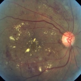
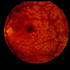
Fundus photograph of left eye of 55-year-old diabetic and hypertensive gentleman with normal serum lipids showing abundant foveal hard exudates.
Condition/keywords: diabetic macular edema
-
---thumb.JPG/image-square;max$300,300.ImageHandler) diabetic macular edema
diabetic macular edema
Oct 26 2012 by Mallika Goyal, MD
Fundus photograph of left eye of 58-year-old diabetic gentleman with normal serum lipids showing foveal hard exudates.
Condition/keywords: foveal hard exudates
-
 Clinically Significant Macular Edema
Apr 23 2015 by Mehul A Shah
Patient presented with complaints of diminished vision ou.
Photographer: Mehul Shah
Imaging device: Zeiss FF450 Plus
Condition/keywords: diabetic macular edema
-
 Vitreoschisis ( Spider-like)
May 31 2014 by Hamid Ahmadieh, MD
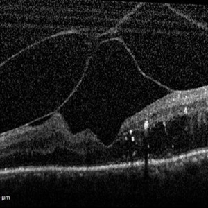
OCT image of the left eye of a 70-year-old woman with proliferative diabetic retinopathy associated with vitreoschisis and diabetic macular edema. The vitreal changes simulate the appearance of a spider on the retina !
Photographer: Nayereh Hadipour, Negah Eye Center, Tehran
Condition/keywords: diabetic macular edema, optical coherence tomography (OCT), vitreoschisis
-
 Fluocinolone Acetonide Intravitreal Implant in Vitreous
Mar 2 2016 by Joshua O Mali, MD, FASRS
Fundus montage displaying Iluvien (fluocinolone acetonide intravitreal implant) in vitreous for treatment of diabetic macular edema.
Condition/keywords: fluocinolone implant, vitreous
-
 Diffuse Diabetic Macular Edema
Sep 8 2012 by Ratimir Lazic, MD, PhD
Color fundus image of a 49 -year - old male. Diffuse macular edema with hard lipid exudates and intraretinal haemorrhages in macular region and mild periphery
Photographer: Ratimir Lazic, PhD MD
Imaging device: Zeis Visucam Lite 2
-
 Focal Laser for CSME
Feb 19 2015 by H. Michael Lambert, MD
Color photo of focal laser near macula.
Condition/keywords: diabetic macular edema, diabetic retinopathy, focal laser
-
 Diabetic Retinopathy Hard Exudates OD
Jun 30 2013 by Rogerio N Shinsato, MD, PhD
Fundus photograph with diabetic retinopathy.
Condition/keywords: diabetic macular edema, foveal hard exudates
-
---thumb.JPG/image-square;max$300,300.ImageHandler) Ischaemic Diabetic Maculopathy
Ischaemic Diabetic Maculopathy
Dec 16 2012 by Mallika Goyal, MD
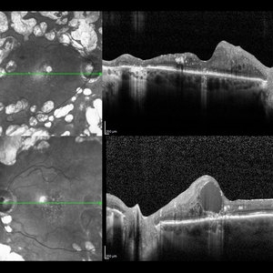
Right eye of a 57 year old diabetic gentleman with diabetic macular edema. OCT shows macular thickening and fluid, and fluorescein shows foveal leak with parafoveal non-perfusion. In view of the parafoveal non-perfusion, anti-VEGF therapy may worsen the situation in this eye.
Photographer: Mallika Goyal, MD, Apollo Health City, Hyderabad, India
Condition/keywords: ischaemic diabetic maculopathy
-
 Diabetic Retinopathy Hard Exudates OD Early Phase
Jun 30 2013 by Rogerio N Shinsato, MD, PhD
Fundus photograph with diabetic retinopathy.
Condition/keywords: diabetic macular edema, foveal hard exudates
-
 Diabetic Retinopathy Hard Exudates OS
Jun 30 2013 by Rogerio N Shinsato, MD, PhD
Fundus photograph with diabetic retinopathy.
Condition/keywords: diabetic macular edema, foveal hard exudates
-
 Diabetic Macular Edema
May 28 2016 by Olivia Rainey
Optical coherence tomography of an 54-year-old female with diabetic macular edema affecting both eyes. Patient has a history of proliferative diabetic retinopathy s/p PRP/PPV/MP/EL, and glaucoma s/p tube shunt in both eyes. There has been a persistence of her macular edema and limited response to antiVEGF therapy, which puts into question whether there is another cause for her edema. Leading the possible causes is her renal insufficiency and fluid retention. Patient was seeing 20/50 in the right eye and 20/80 in the left eye.
Photographer: Olivia Rainey
Imaging device: Heidelberg Spectralis
Condition/keywords: anti-VEGF, diabetic macular edema, edema, glaucoma, optical coherence tomography (OCT), pan-retinal photocoagulation (PRP), proliferative diabetic retinopathy (PDR)
-
 Fluorescein Angiogram - Tortuous Vessels of DME Right Eye
Dec 10 2015 by James B. Soque, CRA, OCT-C, COA, FOPS
Early fluorescein angiogram of diabetic macular edema and tortuous vessels in the superior macula of the right eye.
Photographer: James B Soque, CRA, COA
Imaging device: Top[con TRC-50 DX with MERGE Winstation V 11.2.0
Condition/keywords: diabetes, diabetic macular edema, microaneurysms, microangiopathy, tortuous vessels
-
 Proliferative Diabetic Retinopathy
Mar 16 2015 by Matt Poe, COA
IVFA of 53-year-old male with Proliferative Diabetic Retinopathy, Diabetic macular edema, and a tractional retinal detachment.
Photographer: Matt Poe, COA. Northwest Arkansas Retina Associates, Springdale, AR.
Imaging device: Heidelberg HRA
Condition/keywords: neovascularization (NV), proliferative diabetic retinopathy (PDR)
-
---thumb.JPG/image-square;max$300,300.ImageHandler) Ischaemic diabetic maculopathy
Ischaemic diabetic maculopathy
Dec 16 2012 by Mallika Goyal, MD
Left eye of a 57 year old diabetic gentleman with diabetic macular edema. OCT shows macular thickening and fluid, and fluorescein shows foveal leak with parafoveal non-perfusion. In view of the parafoveal non-perfusion, anti-VEGF therapy may worsen the situation in this eye.
Photographer: Mallika Goyal, MD, Apollo Health City, Hyderabad, India
Condition/keywords: ischaemic diabetic maculopathy
-
 Ozurdex Sarcophagus in DME
Apr 17 2017 by Manish Nagpal, MD, FRCS (UK), FASRS
50-year-old male treated with Ozurdex implant for DME came for a follow up after 4 months and we could see the sarcophagus of the implant dangling in vitreous in front of the macula.
Photographer: POOJA BAROT
Condition/keywords: diabetic macular edema, Ozurdex implant, sarcophagus
-
 Diabetic Macular Edema - Leaking Microaneurysms
Oct 3 2013 by Gerardo Garcia-Aguirre, MD
Diabetic macular edema - leaking microaneurysms.
Condition/keywords: diabetic macular edema, diabetic retinopathy circinate
-
---thumb.JPG/image-square;max$300,300.ImageHandler) Ischaemic diabetic maculopathy
Ischaemic diabetic maculopathy
Dec 16 2012 by Mallika Goyal, MD
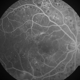
Fluorescein angiogram of right eye of a 57 year old diabetic gentleman with diabetic macular edema reveals significant retinal non-perfusion in the foveal and parafoveal regions. Anti-VEGF therapy may worsen the situation in this eye.
Photographer: Mallika Goyal, MD, Apollo Health City, Hyderabad, India
Condition/keywords: ischaemic diabetic maculopathy
-
 Ozurdex Sarcophagus in DME
Apr 17 2017 by Manish Nagpal, MD, FRCS (UK), FASRS
50-year-old male treated with Ozurdex implant for DME came for a follow up after 4 months and we could see the sarcophagus of the implant dangling in vitreous in front of the macula in this montage view.
Photographer: pooja barot
Condition/keywords: diabetic macular edema, Ozurdex implant, sarcophagus
-
 Ozurdex Sarcophagus in DME
Apr 17 2017 by Manish Nagpal, MD, FRCS (UK), FASRS
50-year-old male treated with Ozurdex implant for DME came for a follow up after 4 months and we could see the sarcophagus of the implant dangling in vitreous in front of the macula.
Photographer: POOJA BAROT
Condition/keywords: diabetic macular edema, Ozurdex implant, sarcophagus
 Loading…