Search results (46 results)
-
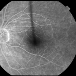
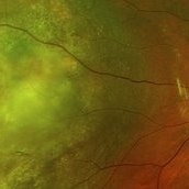
 Orange Pigment Overlying a Lesion Suspicious for a Choroidal Melanoma
Jan 16 2019 by John S. King, MD
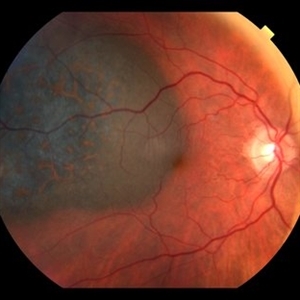
76-year-old white male saw his eye doctor with a three week complaint of photopsias and a shadow in his vision. Found to have a 10.5/12.5/2.5 (medium reflectivity) pigmented, choroidal mass associated with SRF and orange pigment (hyper-autofluorescence of lipofuscin), and without drusen or halo. See photo
Photographer: Stacey Coleman
Imaging device: Topcon 50
Condition/keywords: lipofuscin, orange pigment
-
 Choroidal Mass
Jul 14 2013 by Jason S. Calhoun
Fundus photo shows yellowish choroidal mass in the left eye.
Photographer: Jason S. Calhoun, Department of Ophthalmology, Mayo Clinic Jacksonville, Florida
Imaging device: TOPCON TRC 50-EX
Condition/keywords: choroidal nevus, indolent choroidal mass
-
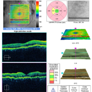
 Choroidal Folds
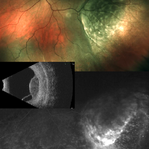
Nov 28 2014 by Thomas A. Ciulla, MD, MBA, FASRS
This 53-year-old man was noted to have choroidal folds right greater than left. The visual acuity was normal at 20/15. The choroidal folds are visible on OCT, especially on the vertical cuts that image across the horizontal folds. Angiography revealed staining of the folds without CNVM, choroidal mass, or optic nerve edema.
Photographer: Charlotte Harris
Condition/keywords: bilateral chorioretinal folds, choroidal folds
-
---thumb.jpg/image-square;max$300,300.ImageHandler) Vitrectomy Choroidal Mass
Vitrectomy Choroidal Mass
Feb 13 2013 by From the Collections of Thomas M. Aaberg, MD and Thomas M. Aaberg Jr., MD
Tumor versus serous choroidal elevation.
Condition/keywords: choroidal mass, tumor, vitrectomy
-
 Choroidal Mass
Sep 21 2018 by Sarah Oelrich
Choroidal mass
Photographer: Sarah Oelrich CRA COT, Southeastern Retina Associates Knoxville Tn
Imaging device: OPTOS 200tx
Condition/keywords: choroidal mass
-
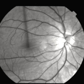
 Peripapillary Choroidal Mass
Oct 30 2015 by Natalie Loyacano, COMT, OCS-R,OSA, ROUB
Fundus photograph of 50 year-old male with a suspicious peripapillary pigmented lesion. Patient sees a spot in his vision that has progressively worsen over the past month.
Photographer: Amy Gunter, VitreoRetinal Eye Center, Biloxi MS
Imaging device: Topcon
Condition/keywords: choroidal mass
-
 Choroidal Folds
Nov 28 2014 by Thomas A. Ciulla, MD, MBA, FASRS
This 53-year-old man was noted to have choroidal folds right greater than left. The visual acuity was normal at 20/15. The choroidal folds are visible on OCT, especially on the vertical cuts that image across the horizontal folds. Angiography revealed staining of the folds without CNVM, choroidal mass, or optic nerve edema.
Photographer: Charlotte Harris
Condition/keywords: bilateral chorioretinal folds, choroidal folds
-
 Choroidal Folds
Nov 28 2014 by Thomas A. Ciulla, MD, MBA, FASRS
This 53 -year-old man was noted to have choroidal folds right greater than left. The visual acuity was normal at 20/15. The choroidal folds are visible on OCT, especially on the vertical cuts that image across the horizontal folds. Angiography revealed staining of the folds without CNVM, choroidal mass, or optic nerve edema.
Condition/keywords: bilateral chorioretinal folds, choroidal folds
-
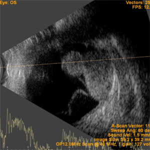 Collar Button Appearance on B-Scan
Aug 28 2019 by Gayathri Mohan
B-scan showing an intraocular mass with collar button appearance. Suspected case of choroidal melanoma.
Photographer: Dr.Gayathri Mohan, Retina Foundation
Imaging device: Nidek Mirante SLO
Condition/keywords: choroidal mass, collar button
-
---thumb.JPG/image-square;max$300,300.ImageHandler) Choroidal Mass 1
Choroidal Mass 1
Jul 14 2013 by Jason S. Calhoun
Fundus photo shows large elevated choroidal melanoma centrally in the left eye.
Photographer: Jason S. Calhoun, Department of Ophthalmology, Mayo Clinic Jacksonville, Florida
Imaging device: TOPCON TRC 50-EX
-
 Choroidal Metastasis With Serous Retinal Detachment Rigth Eye
Sep 2 2015 by María José Marroquín Sarti
A 62-year-old woman complained of visual field loss and decreasing vision. Twenty years earlier, breast cancer was diagnosed and treated with chemotherapy and right mastectomy, four years ago, she had another treatment with chemotherapy and resection of another tumor that started to grow in the same side. Serous retinal detachment and choroidal masses were present in both eyes.
Photographer: María José Marroquín Sarti, Escuela Superior de Oftalmología
Condition/keywords: bilateral serous detachment, choroidal metastasis
-
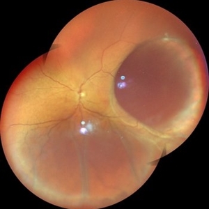
 Wide Field Fundus Montage of Intraocular Mass with Retinal Detachment
Aug 28 2019 by Gayathri Mohan
50 year old female came with diminution of vision in the LE. Wide field fundus photograph showing an intraocular mass temporally along with an exudative retinal detachment inferiorly. Ultrasonography showed an intraocular mass with collar button appearance suggestive of a Choroidal melanoma. She underwent enucleation and histopathology confirmed a spindle cell choroidal melanoma
Photographer: Dr. Gayathri Mohan, Retina Foundation
Imaging device: Nidek Mirante SLO
Condition/keywords: choroidal mass, collar button
-
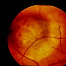
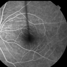
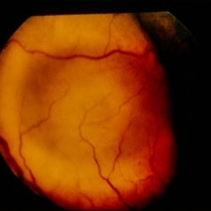 Amelanotic Choroidal Melanoma
Jan 29 2015 by H. Michael Lambert, MD
Elevated choroidal mass.
-
 Intraocular Mass With Retinal Detachment
Aug 28 2019 by Gayathri Mohan
Wide field fundus image showing an intraocular mass temporally along with a retinal detachment.
Photographer: Dr. Gayathri Mohan, Retina Foundation
Imaging device: Nidek Mirante SLO
Condition/keywords: choroidal mass
-
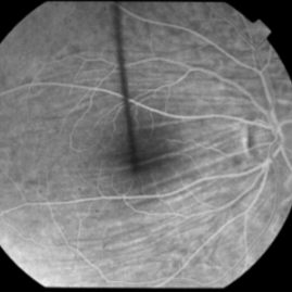
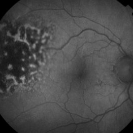 Hyper-autofluorescence of Orange Pigment Overlying a Lesion Suspicious for a Choroidal Melanoma
Jan 16 2019 by John S. King, MD
76-year-old white male saw his eye doctor with a three week complaint of photopsias and a shadow in his vision. Found to have a 10.5/12.5/2.5 (medium reflectivity) pigmented, choroidal mass associated with SRF and orange pigment (hyper-autofluorescence of lipofuscin, see image), and without drusen or halo.
Photographer: Stacey Coleman
Imaging device: Topcon 50
Condition/keywords: lipofuscin, orange pigment
-
 Choroidal Folds
Nov 28 2014 by Thomas A. Ciulla, MD, MBA, FASRS
This 53-year-old man was noted to have choroidal folds right greater than left. The visual acuity was normal at 20/15. The choroidal folds are visible on OCT, especially on the vertical cuts that image across the horizontal folds. Angiography revealed staining of the folds without CNVM, choroidal mass, or optic nerve edema.
Photographer: Charlotte Harris
Condition/keywords: bilateral chorioretinal folds, choroidal folds
-
 Amelanotic Choroidal Melanoma
Jan 29 2015 by H. Michael Lambert, MD
Elevated choroidal mass.
-
 Choroidal Folds
Nov 28 2014 by Thomas A. Ciulla, MD, MBA, FASRS
This 53-year-old man was noted to have choroidal folds right greater than left. The visual acuity was normal at 20/15. The choroidal folds are visible on OCT, especially on the vertical cuts that image across the horizontal folds. Angiography revealed staining of the folds without CNVM, choroidal mass, or optic nerve edema.
Condition/keywords: bilateral chorioretinal folds, choroidal folds
-
 Choroidal Folds
Nov 28 2014 by Thomas A. Ciulla, MD, MBA, FASRS
This 53-year-old man was noted to have choroidal folds right greater than left. The visual acuity was normal at 20/15. The choroidal folds are visible on OCT, especially on the vertical cuts that image across the horizontal folds. Angiography revealed staining of the folds without CNVM, choroidal mass, or optic nerve edema.
Photographer: Charlotte Harris
Condition/keywords: bilateral chorioretinal folds, choroidal folds
-
 Choroidal-Mass with Exudative Retinal Detachment
Nov 23 2021 by VIRAL SHAH
48 year-old male patient has complaint of dimness of vision in left eye for 1-1/2 months. He has history of Radical Nephrectomy of left side due to clear cell renal cell carcinoma 4 months back.
Photographer: VIRAL SHAH, NETRALOK RETINA CLINIC, AHMEDABAD
Condition/keywords: choroidal mass, unilateral exudative retinal detachment
-
 Choroidal Melanoma with Exudative Retinal Detachment
Mar 2 2023 by Aditya S Kelkar, MS, FRCS, FASRS,FRCOphth
Color fundus photograph of the left eye of a 45 year old male showing choroidal melanoma with exudative retinal detachment.
Photographer: Dr. Pranali Surawase, National Institute of Ophthalmology, Pune, India.
Imaging device: Zeiss Clarus 500
Condition/keywords: choroidal mass, exudative retinal detachment, Retinal detachment
-
 Choroidal Folds
Nov 28 2014 by Thomas A. Ciulla, MD, MBA, FASRS
This 53-year-old man was noted to have choroidal folds right greater than left. The visual acuity was normal at 20/15. The choroidal folds are visible on OCT, especially on the vertical cuts that image across the horizontal folds. Angiography revealed staining of the folds without CNVM, choroidal mass, or optic nerve edema.
Photographer: Charlotte Harris
Condition/keywords: bilateral chorioretinal folds, choroidal folds
-
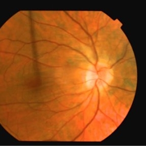
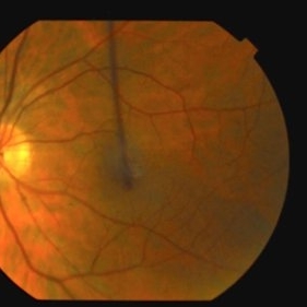
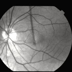
 Nasal Choroidal Mass
Jan 7 2018 by John S. King, MD
Mets vs Melanoma
Imaging device: Optos
Condition/keywords: choroidal mass
 Loading…