Search results (87 results)
-
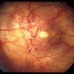
 Myopic macular degeneration
Jan 11 2013 by Alex P. Hunyor, MD
Myopic macular degeneration, left eye - extensive chorioretinal atrophy.
Condition/keywords: myopic degeneration, myopic fundus, myopic macular degeneration
-
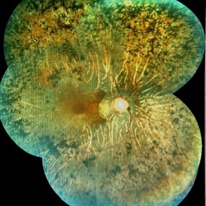
 High Myopia
May 2 2013 by Henry J. Kaplan, MD
Chorioretinal atrophy in high myopia and tilted disc.
Condition/keywords: high myopia, tilted disc
-
---thumb.jpg/image-square;max$300,300.ImageHandler) Posterior Pole Chorioretinal Atrophy With Staphyloma
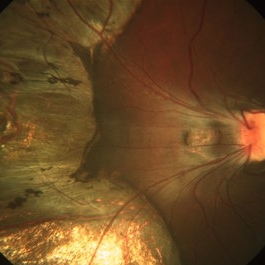
Posterior Pole Chorioretinal Atrophy With Staphyloma
Aug 1 2013 by From the Collections of Thomas M. Aaberg, MD and Thomas M. Aaberg Jr., MD
Posterior pole chorioretinal atrophy with staphyloma.
Condition/keywords: chorioretinal atrophy, posterior pole, staphyloma
-
---thumb.jpg/image-square;max$300,300.ImageHandler) Posterior Pole Chorioretinal Atrophy With Staphyloma
Posterior Pole Chorioretinal Atrophy With Staphyloma
Aug 1 2013 by From the Collections of Thomas M. Aaberg, MD and Thomas M. Aaberg Jr., MD
Posterior pole chorioretinal atrophy with staphyloma.
Condition/keywords: chorioretinal atrophy, staphyloma
-
 Choroideremia
Jan 26 2013 by Ratimir Lazic, MD, PhD
FAG image of a 66-year-old male. Diffuse chorioretinal atrophy is present. Large choroidal vessels can be seen due to atrophy of the RPE and choriocapilaris.
Photographer: Marko Lukic, MD
Imaging device: Zeis Visucam Lite 2
Condition/keywords: choroideremia, fundus photograph
-
 Choroideremia
Jan 26 2013 by Ratimir Lazic, MD, PhD
Color fundus photography of a 66-year-old male. Diffuse chorioretinal atrophy is seen. “Patches” or retinal tissue can be seen, some of it in macular area. Visual acuity on that eye is 0,15.
Photographer: Marko Lukic, MD
Imaging device: Zeis Visucam Lite 2
Condition/keywords: choroideremia, large choroidal vessels
-
 Choroideremia
Jan 26 2013 by Ratimir Lazic, MD, PhD
FAG image of a 66-year-old male. Diffuse chorioretinal atrophy is present. Large choroidal vessels can be seen. "Hyperflorescent" areas represent normal chorioretinal tissue.
Photographer: Marko Lukic, MD
Imaging device: Zeis Visucam Lite 2
Condition/keywords: choroideremia, fundus photograph
-
 Progressive Bifocal Chorioretinal Atrophy
Feb 1 2015 by Andree Henaine-Berra, MD
Fundus photograph of the left eye of an 13-year-old female patient with poor vision, high myopia and nystagmus. The image shows macular dragging, a limited area of chorioretinal atrophy temporal to the optic disc and an extense area of chorioretinal atrophy temporal to the macula that extended to the extreme periphery.
Photographer: Andree Henaine-Berra, MD
Condition/keywords: chorioretinal atrophy
-
 Colour Photo of a Rare Case of Pigmented Paravenous Chorioretinal Atrophy (PPCRA)
Jun 30 2017 by Manish Nagpal, MD, FRCS (UK), FASRS
A rare case of PPCRA
Photographer: RAKESH JUNEJA
Condition/keywords: pigmented paravenous chorioretinal atrophy (PPCRA), rare
-
 Colour Photo of a Case of Pigmented Paravenous Chorioretinal Atrophy (PPCRA)
Jun 30 2017 by Manish Nagpal, MD, FRCS (UK), FASRS
This is a rare presentation of a case of PPCRA
Photographer: RAKESH JUNEJA
Condition/keywords: pigmented paravenous chorioretinal atrophy (PPCRA), rare
-
---thumb.jpg/image-square;max$300,300.ImageHandler) Posterior Pole Chorioretinal Atrophy With Staphyloma
Posterior Pole Chorioretinal Atrophy With Staphyloma
Aug 1 2013 by From the Collections of Thomas M. Aaberg, MD and Thomas M. Aaberg Jr., MD
Posterior pole chorioretinal atrophy with staphyloma.
Condition/keywords: chorioretinal atrophy, posterior pole, staphyloma
-
 High Myopia
Jun 14 2018 by Mitzy E Torres Soriano, MD
Fundus photograph (left eye) of a female patient with high myopia, chorioretinal atrophy, pigmentary changes and posterior staphyloma.
Photographer: Mitzy Torres Soriano
Condition/keywords: chorioretinal atrophy, high myopia, posterior staphyloma
-
---thumb.JPG/image-square;max$300,300.ImageHandler) choroidal lymphoma
choroidal lymphoma
Nov 25 2012 by Mallika Goyal, MD
Left eye of a 60-year-old lady shows areas of chorio-retinal atrophy corresponding to regression of choroidal lymphoma following external beam irradiation.
Photographer: Mallika Goyal, MD, Apollo Health City, Hyderabad, India
Condition/keywords: chorioretinal atrophy, lymphoma
-
---thumb.jpg/image-square;max$300,300.ImageHandler) Posterior Pole Chorioretinal Atrophy With Staphyloma
Posterior Pole Chorioretinal Atrophy With Staphyloma
Aug 1 2013 by From the Collections of Thomas M. Aaberg, MD and Thomas M. Aaberg Jr., MD
Posterior pole chorioretinal atrophy with staphyloma.
Condition/keywords: chorioretinal atrophy, staphyloma
-
 OCT OS of a Case of Pigmented Paravenous Chorioretinal Atrophy (PPCRA)
Jun 30 2017 by Manish Nagpal, MD, FRCS (UK), FASRS
This is the OCT of a rare case of PPCRA.
Photographer: Rakesh Junena
Condition/keywords: pigmented paravenous chorioretinal atrophy (PPCRA), rare
-
 OCT OD of a Case of Pigmented Paravenous Chorioretinal Atrophy (PPCRA)
Jun 30 2017 by Manish Nagpal, MD, FRCS (UK), FASRS
This is the OCT of a rare case of PPCRA
Photographer: RAKESH JUNEJA
Condition/keywords: pigmented paravenous chorioretinal atrophy (PPCRA), rare
-
 Retinitis Pigmentosa
Feb 26 2020 by Manuel Ángel Alcántara Delgado, MD
Merged color fundus photograph of a 68-year-old woman with advanced retinitis pigmentosa. It is appreciated bone spicule-shaped pigment deposits, optic disc pallor, retinal atrophy, attenuated retinal vessels and surface wrinkling retinopathy.
Photographer: Manuel Ángel Alcántara Delgado
Condition/keywords: chorioretinal atrophy, choroidal circulation, optic disc pallor, pericentral retinitis pigmentosa, retina, retinitis pigmentosa, retinitis pigmentosa (RP) dystrophy, sector retinitis pigmentosa
-
 Pigmented Paravenous Chorioretinal Atrophy
Feb 1 2018 by John S. King, MD
15-year-old healthy, asymptomatic AAM; found on routine eye exam; no FHx of RP known; OCT shows some extrafoveal, outer retinal degeneration.
Photographer: Karin Aletter
Condition/keywords: pigmented paravenous chorioretinal atrophy (PPCRA)
-
 Toxoplasmosis
Feb 13 2013 by From the Collections of Thomas M. Aaberg, MD and Thomas M. Aaberg Jr., MD
Chorioretinal scar, chorioretinal atrophy.
Condition/keywords: chorioretinal atrophy, chorioretinal scar, toxoplasmosis
-
 Pigmented Paravenous Chorioretinal Atrophy
Feb 1 2018 by John S. King, MD
15 yo healthy, asymptomatic AAM; found on routine eye exam; no FHx of RP known; OCT shows some extrafoveal, outer retinal degeneration.
Photographer: Karin Aletter
Condition/keywords: pigmented paravenous chorioretinal atrophy (PPCRA)
-
 Progressive Bifocal Chorioretinal Atrophy
Feb 1 2015 by Andree Henaine-Berra, MD
Fundus photograph of the right eye of an 13-year-old female patient with poor vision, high myopia and nystagmus. The image shows macular dragging, a limited area of chorioretinal atrophy temporal to the optic disc and an extense area of chorioretinal atrophy temporal to the macula that extended to the extreme periphery.
Photographer: Andree Henaine-Berra, MD
Condition/keywords: chorioretinal atrophy
-
 Traumatic Optic Neuropathy With Macular Scar
May 4 2014 by Mallika Goyal, MD
Left eye of a 36-year-old male patient reveals extensive chorioretinal atrophy in the inferior mid-periphery and periphery 4 weeks after blunt injury with left orbital fracture in a road accident. The atrophy is likely secondary to gravitational inferior tracking of massive submacular bleed during the concussion.
Photographer: Mallika Goyal, MD, Apollo Health City, Jubilee Hills, Hyderabad, India
Condition/keywords: traumatic optic neuropathy
-
 Pigmented Paravenous Chorioretinal Atrophy
Feb 1 2018 by John S. King, MD
15-year-old healthy, asymptomatic AAM; found on routine eye exam; no FHx of RP known; OCT shows some extrafoveal, outer retinal degeneration.
Photographer: Karin Aletter
Condition/keywords: pigmented paravenous chorioretinal atrophy (PPCRA)
-
 Case of Pigmented Para Venous Chorioretinal Atrophy in a Girl Marfan
Jul 11 2013 by Eric M. Shrier, DO
14-year-old black female with Marfan Syndrome.
Condition/keywords: choroidal atrophy
 Loading…