Search results (252 results)
-
 PED due to CSCR
Sep 2 2012 by Hamid Ahmadieh, MD
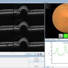
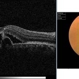
OCT image of a 37-year-old man with a serous PED secondary to CSCR.
Photographer: Hamid Ahmadieh, Ophthalmic Research Center, Labbafinejad Medical Center
Imaging device: Heidelberg Spectralis
Condition/keywords: central serous chorioretinopathy (CSCR), optical coherence tomography (OCT), pigment epithelial detachment (PED)
-
 Chronic Active Central Serous Chorioretinopathy (CSCR)
Sep 11 2012 by Hamid Ahmadieh, MD
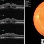
Color fundus photograph and OCT image of a 30-year-old man with chronic active CSCR.
Photographer: Hamid Ahmadieh, MD, Ophthalmic Research Center, Labbafinejad Medical Center, Shahid Beheshti University of Medical Sciences
Imaging device: Topcon
Condition/keywords: central serous chorioretinopathy (CSCR), optical coherence tomography (OCT)
-
 Recurrent Central Serous Choroidopathy
Aug 21 2012 by Edwin H. Ryan, MD
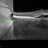
EDI-OCT showing thickened choroid and subretinal fluid
Photographer: Edwin Ryan Jr. MD, VitreoRetinal Surgery, PA
Imaging device: Heidelberg Spectralis
Condition/keywords: central serous chorioretinopathy (CSCR), choroidal thickening, enhanced depth imaging
-
 Multifocal CSCR 2
Sep 2 2012 by Hamid Ahmadieh, MD
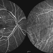
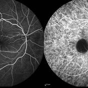
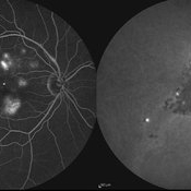
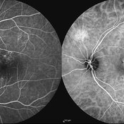
Early-phase FA and ICG angiograms of a 36-year-old man with an active multifocal CSCR.
Photographer: Hamid Ahmadieh, Ophthalmic Research Center, Labbafinejad Medical Center
Imaging device: Heidelberg Spectralis
Condition/keywords: central serous chorioretinopathy (CSCR), indocyanine green (ICG) angiography
-
 Central Serous Chorioretinopathy, Fluorescein Angiogram
Aug 23 2012 by Gerardo Garcia-Aguirre, MD
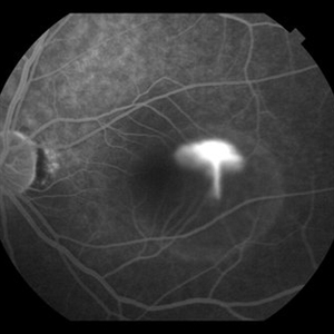
Fluorescein angiogram, late phase, showing hyperfluorescent spot, larger than earlier phases.
Photographer: Noemí Hernández, Asociación para Evitar la Ceguera en México
Imaging device: Zeiss FF4
Condition/keywords: central serous chorioretinopathy (CSCR)
-
 PED due to CSCR 4
Sep 2 2012 by Hamid Ahmadieh, MD
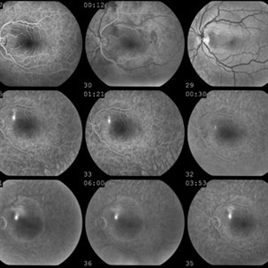
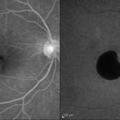
Early phase FA & ICG images of a 37-year-old man with a serous PED secondary to CSCR
Photographer: Hamid Ahmadieh, Ophthalmic Research Center, Labbafinejad Medical Center
Imaging device: Heidelberg Spectralis
Condition/keywords: central serous chorioretinopathy (CSCR), indocyanine green (ICG) angiography, pigment epithelial detachment (PED)
-
 PED due to CSCR 2
Sep 2 2012 by Hamid Ahmadieh, MD
Autofluorescence imaging of a 37-year-old man with a serous PED secondary to CSCR.
Photographer: Hamid Ahmadieh, Ophthalmic Research Center, Labbafinejad Medical Center
Imaging device: Heidelberg Spectralis
Condition/keywords: autofluorescence imaging, central serous chorioretinopathy (CSCR), pigment epithelial detachment (PED)
-
 Smokestack Sign
Oct 13 2012 by Geoffrey G. Emerson, MD, PhD, FASRS
Central serous choroidopathy, smokestack
Condition/keywords: central serous chorioretinopathy (CSCR)
-
 "Smoke Stack" Hyperfluorescence in Central Serous Chorioretinopathy
Mar 2 2014 by Homayoun Tabandeh, MD, FASRS
"Smoke Stack" hyperfluorescence in central serous chorioretinopathy.
Condition/keywords: central serous chorioretinopathy (CSCR)
-
 Multifocal CSCR 1
Sep 2 2012 by Hamid Ahmadieh, MD
Fundus autofluorescence of a 36-year-old man with an active multifocal CSCR.
Photographer: Hamid Ahmadieh, Ophthalmic Research Center, Labbafinejad Medical Center
Imaging device: Heidelberg Spectralis
Condition/keywords: central serous chorioretinopathy (CSCR)
-
 Central Serous Chorioretinopathy
Aug 23 2012 by Gerardo Garcia-Aguirre, MD
Fundus photograph of a 29 year-old patient showing subretinal fluid in the macula.
Photographer: Noemí Hernández, Asociación para Evitar la Ceguera en México
Imaging device: Zeiss FF4
Condition/keywords: central serous chorioretinopathy (CSCR)
-
 Recurrent Central Serous Chorioretinopathy
Aug 22 2012 by Edwin H. Ryan, MD
fundus fluorescein angiogram of 55 year-old man with recurrent central serous, with fibrin
Photographer: Edwin Ryan Jr. MD, VitreoRetinal Surgery, PA
Condition/keywords: central serous chorioretinopathy (CSCR), subretinal fibrin deposition
-
 Central Serous Chorioretinopathy (CSC)
Oct 16 2012 by S. Natarajan, MD, FASRS, FRCS (GLASGOW) , FICO, D.Sc, FELA
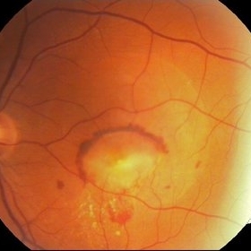
Middle-aged male came with small PED 4 months back; now this has progressed to a larger PED with SRF underneath the fovea.
Photographer: Prof. Dr. S. Natarajan
Condition/keywords: central serous chorioretinopathy (CSCR), central serous retinopathy (CSR), pigment epithelial detachment (PED), subretinal fibrosis
-
 Multifocal CSCR
Sep 2 2012 by Hamid Ahmadieh, MD
Late-phase FA and ICG angiograms of a 36-year-old man with an active multifocal CSCR.
Photographer: Hamid Ahmadieh, Ophthalmic Research Center, Labbafinejad Medical Center
Imaging device: Heidelberg Spectralis
Condition/keywords: central serous chorioretinopathy (CSCR), indocyanine green (ICG) angiography
-
 Recurrent Central Serous Chorioretinopathy
Aug 22 2012 by Edwin H. Ryan, MD
Fundus fluorescein angiogram of 55-year-old man with recurrent central serous, with fibrin.
Photographer: Edwin Ryan Jr. MD, VitreoRetinal Surgery, PA
Condition/keywords: central serous chorioretinopathy (CSCR), fluorescein leakage
-
 Chronic Central Serous Chorioretinopathy
Sep 26 2012 by Hamid Ahmadieh, MD
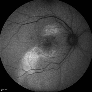
Autofluorescence imaging of the right eye of a 50-year-old man with active chronic CSCR and BCVA of 20/100.
Photographer: Hamid Ahmadieh, MD, Ophthalmic Research Center, Labbafinejad Medical Center, Shahid Beheshti University of Medical Sciences
Imaging device: Heidelberg Spectralis
Condition/keywords: autofluorescence imaging, chronic central serous chorioretinopathy (CSCR)
-
 Central Serous Chorioretinopathy Smokestack 4
Aug 24 2012 by John S. King, MD
Smokestack
Photographer: Kristin Konecki, OcuSight Eye Care Center, Rochester, NY
Condition/keywords: central serous chorioretinopathy (CSCR), smokestack
-
 Chronic Central Serous Chorioretinopathy
Sep 26 2012 by Hamid Ahmadieh, MD
Color fundus photograph and OCT imaging of the right eye of a 50-year-old man with active chronic CSCR and BCVA of 20/100
Photographer: Hamid Ahmadieh, MD, Ophthalmic Research Center, Labbafinejad Medical Center, Shahid Beheshti University of Medical Sciences
Imaging device: Topcon OCT
Condition/keywords: chronic central serous chorioretinopathy (CSCR), optical coherence tomography (OCT)
-
 Central Serous Chorioretinopathy, Fluorescein Angiogram
Aug 23 2012 by Gerardo Garcia-Aguirre, MD
Fluorescein angiogram, late phase, showing a small, hyperfluorescent spot.
Photographer: Noemí Hernández, Asociación para Evitar la Ceguera en México
Imaging device: Zeiss FF4
Condition/keywords: central serous chorioretinopathy (CSCR)
-
 PED due to CSCR 5
Sep 2 2012 by Hamid Ahmadieh, MD
Late-phase FA and ICG images of a 37-year-old man with a serous PED secondary to CSCR.
Photographer: Hamid Ahmadieh, Ophthalmic Research Center, Labbafinejad Medical Center
Imaging device: Heidelberg Spectralis
Condition/keywords: central serous chorioretinopathy (CSCR), indocyanine green (ICG) angiography, pigment epithelial detachment (PED)
-
 Chronic Active Central Serous Chorioretinopathy (CSCR)
Sep 11 2012 by Hamid Ahmadieh, MD
Late phase FA & ICG angiography images of a 30-year-old man with chronic active CSCR.
Photographer: Hamid Ahmadieh, MD, Ophthalmic Research Center, Labbafinejad Medical Center, Shahid Beheshti University of Medical Sciences
Imaging device: Heidelberg Spectralis
Condition/keywords: central serous chorioretinopathy (CSCR), indocyanine green (ICG) angiography
-
 PED due to CSCR 3
Sep 2 2012 by Hamid Ahmadieh, MD
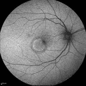
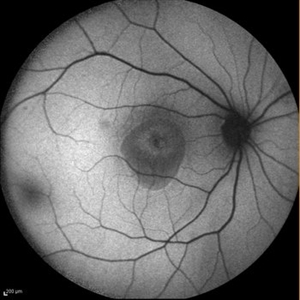
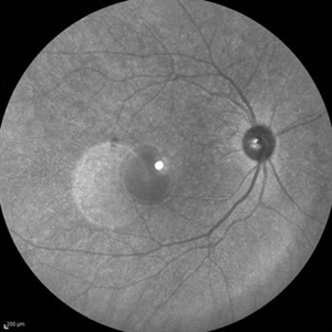
Infrared imaging of a 37-year-old man with a serous PED secondary to CSCR.
Photographer: Hamid Ahmadieh, Ophthalmic Research Center, Labbafinejad Medical Center
Imaging device: Heidelberg Spectralis
Condition/keywords: central serous chorioretinopathy (CSCR), pigment epithelial detachment (PED)
-
 Central Serous Chorioretinopathy Smokestack 3
Aug 24 2012 by John S. King, MD
Smokestack
Photographer: Kristin Konecki, OcuSight Eye Care Center, Rochester, NY
Condition/keywords: central serous chorioretinopathy (CSCR), smokestack
-
---thumb.jpg/image-square;max$300,300.ImageHandler) Central Serous Chorioretinopathy 2
Central Serous Chorioretinopathy 2
Mar 18 2013 by Maurice F. Rabb
Woman with a 3 month history of reduced vision, and her fundi appeared as if she had a severe form of central serous chorioretinopathy, including subretinal febrin deposition, serous pigment epithelial detachments, patchy zones of pigment epithelial atrophy, and dependent, bullous detachments bilaterally. There are also multifocal areas of orange subretinal deposits, some in the form of an irregular sequence or change. These looked like Elschnig spots and Siegrist lines, consistent with choroidal ischemia that could account for the exudative detachments as well.
Condition/keywords: bullous detachments bilaterally, central serous chorioretinopathy (CSCR), choroidal ischemia, dependent, orange subretinal deposits, patchy zones of pigment epithelial atrophy, reduced vision, serous pigment epithelial detachment, Siegrist Streaks, subretinal fibrin deposition
-
 CNV Due to Chronic Central Serous Retinopathy
Apr 6 2014 by Ratimir Lazic, MD, PhD
A color fundus image of a 66-year-old male with previously diagnosed chronic CSR. Few weeks ago the patient noticed rapidly worsening of the visual acuity on the left eye. Subretinal hemorrhage with big PED and subretinal exudation can be noticed. The image presents baseline clinical picture of the left eye. The antiVEGF intravitreal treatment have been started.
Photographer: Marko Lukic, University Eye Clinic Svjetlost
Imaging device: Zeis Visucam Lite 2
Condition/keywords: central serous chorioretinopathy (CSCR), choroidal neovascularization (CNV), subretinal hemorrhage
 Loading…