Search results (261 results)
-
 CRVO with Flame Hemorrhages
Oct 1 2012 by Jeffrey G. Gross, MD, FASRS
CRVO with flame hemorrhages and cotton wool spots 20/80.
Condition/keywords: 20/80, central retinal vein occlusion (CRVO), cotton wool spots
-
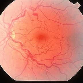
 Optociliary Shunt Vessels in Old CRVO
Sep 8 2012 by Hamid Ahmadieh, MD
FA image of a 60-year-old woman with the history of central retinal vein occlusion.
Photographer: Hamid Ahmadieh, MD, Ophthalmic Research Center, Labbafinejad Medical Center, Shahid Beheshti University of Medical Sciences
Imaging device: Heidelberg Spectralis
Condition/keywords: central retinal vein occlusion (CRVO), shunts vessels
-
 Non Ischemic Hemi-CRVO
Mar 29 2013 by Henry J. Kaplan, MD
Non-ischemic CRVO: blurred disc margins, dilated and tortous veins and scattered hemorrhages in the superior half of the retina.
Condition/keywords: branch retinal vein occlusion (BRVO), central retinal vein occlusion (CRVO), hemi CRVO, non-ischemic central retinal vein occlusion (CRVO)
-
 Non Ischemic CRVO
Mar 29 2013 by Henry J. Kaplan, MD
Non ischemic CRVO.
Condition/keywords: central retinal vein occlusion (CRVO)
-
 Rubeosis and Fibrin
Oct 1 2012 by Jeffrey G. Gross, MD, FASRS
Rubeosis and fibrin in patient with CRVO CF.
Condition/keywords: central retinal vein occlusion (CRVO), fibrin, rubeosis
-
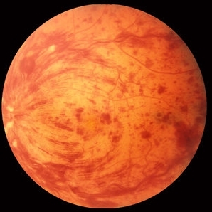
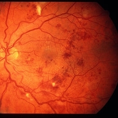
 Central Retinal Vein Occlusion
Sep 2 2012 by Hyung-Woo Kwak, MD
Multiple dense, dark, blotchy hemorrhages, cotton-wool spots, and pale optic disc are signs suggestive of retinal ischemia in CRVO.
Imaging device: Zeiss F450 plus
Condition/keywords: central retinal vein occlusion (CRVO)
-
---thumb.jpg/image-square;max$300,300.ImageHandler) Cilioretinal Artery Occlusion with Central Retinal Vein Occlusion
Cilioretinal Artery Occlusion with Central Retinal Vein Occlusion
Mar 9 2013 by Gabriela Lopezcarasa Hernandez, MD
A 46-year-old male with decrease in visual acuity in left eye and central scotoma.
Photographer: Araceli Rojas Arriaga, Hospital Angeles Lomas, Mexico
Imaging device: Zeiss FF4
Condition/keywords: central retinal vein occlusion (CRVO), cilioretinal artery occlusion
-
 Venous Beading
Nov 4 2021 by Stefanie Palmer
Venous Beading in a patient with both PDR and CRVO.
Photographer: Stefanie Palmer, CRA
Imaging device: Topcon
Condition/keywords: central retinal vein occlusion (CRVO), diabetic retinopathy, proliferative diabetic retinopathy (PDR), venous beading
-
 Impending CRVO
Mar 29 2013 by Henry J. Kaplan, MD
Dilated and tortous retinal veins and scant hemorrhage in impending CRVO.
Condition/keywords: central retinal vein occlusion (CRVO)
-
 Polycythemia Vera
May 2 2013 by Henry J. Kaplan, MD
CRVO in polycythemia vera; dilated and tortous retinal veins, hemorrhages and cotton wool spots.
Condition/keywords: central retinal vein occlusion (CRVO), polycythemia vera
-
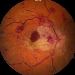
---thumb.jpg/image-square;max$300,300.ImageHandler) Central Retinal Vein Occlusion
Central Retinal Vein Occlusion
Oct 30 2012 by Lihteh Wu, MD
35-year-old hypertensive man with an acute CRVO. Notice the peripapillary cotton wool spots, superficial flame shaped hemorrhages and deeper dot and blot hemorrhages in all 4 quadrants. This is the typical blood and thunder appearance of a CRVO.
Condition/keywords: central retinal vein occlusion (CRVO), cotton wool spots
-
 Branch Retinal Artery Occlusion With Concurrent Central Retinal Vein Occlusion
Oct 5 2016 by Larry M Puthenparambil, MD
Branch retinal artery occlusion with concurrent central retinal vein occlusion.
Photographer: Stacey Groom
Imaging device: Topcon
Condition/keywords: branch retinal artery occlusion (BRAO), central retinal vein occlusion (CRVO)
-
---thumb.jpg/image-square;max$300,300.ImageHandler) Cilioretinal Artery Occlusion with Central Retinal Vein Occlusion
Cilioretinal Artery Occlusion with Central Retinal Vein Occlusion
Mar 9 2013 by Gabriela Lopezcarasa Hernandez, MD
A 46-year-old male with decrease in visual acuity in left eye and central scotoma.
Photographer: Araceli Rojas Arriaga, Hospital Angeles Lomas, Mexico
Imaging device: Zeiss FF4
Condition/keywords: central retinal artery occlusion (CRAO), central retinal vein occlusion (CRVO), cilioretinal artery occlusion
-
 Impending CRVO
Mar 29 2013 by Henry J. Kaplan, MD
Venous dilation and torousity and faint spot hemorrhages in a patient with impending CRVO.
Condition/keywords: central retinal vein occlusion (CRVO)
-
 CRVO Colour
Oct 8 2012 by David R. Chow, MD, FRCS(C)
Condition/keywords: central retinal vein occlusion (CRVO)
-
 CRVO with cilioretinal artery occlusion
Jan 11 2013 by Alex P. Hunyor, MD
Nonischaemic central retinal vein obstruction (CRVO) with cilioretinal artery occlusion.
Condition/keywords: central retinal vein occlusion (CRVO), cilioretinal artery occlusion
-
 CRVO-associated Macular Edema
Jul 8 2012 by Jeffrey S. Heier, MD
Young physician with CRVO and macular edema
Imaging device: Spectralis
Condition/keywords: central retinal vein occlusion (CRVO), macular edema
-
---thumb.jpg/image-square;max$300,300.ImageHandler) Central Retinal Vein Occlusion
Central Retinal Vein Occlusion
Oct 30 2012 by Lihteh Wu, MD
35-year-old hypertensive man with an acute CRVO. Notice the peripapillary cotton wool spots, superficial flame shaped hemorrhages and deeper dot and blot hemorrhages in all 4 quadrants. This is the typical blood and thunder appearance of a CRVO.
Condition/keywords: central retinal vein occlusion (CRVO), cotton wool spots
-
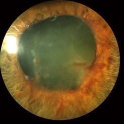
 Combined CRAO and CRVO
May 15 2014 by Manish Nagpal, MD, FRCS (UK), FASRS
30-year-old anemic lady presented with a acute loss of vision. Her vision was just hand movements in the affected eye and the other eye was normal.
Photographer: pooja barot, Optometrist, Retina Foundation, Ahmedabad
Condition/keywords: central retinal artery occlusion (CRAO), central retinal vein occlusion (CRVO), macular edema
-
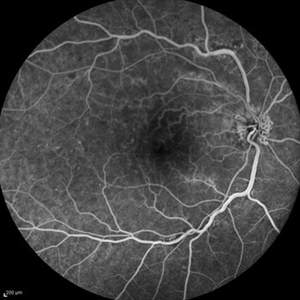
 Central Retinal Vein Occlusion: Case 1
Oct 12 2012 by Gregg T. Kokame, MD, MMM, FASRS
Early-phase FA
Photographer: Jaclyn Pisano, Retina Consultants of Hawaii
Imaging device: Topcon 50IA / Eschalon
Condition/keywords: central retinal vein occlusion (CRVO)
-
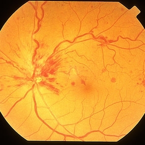
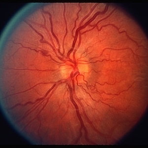
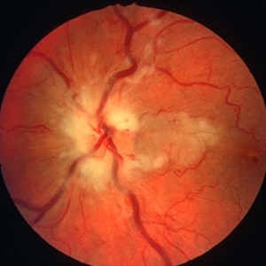
---thumb.JPG/image-square;max$300,300.ImageHandler) Disc Collaterals
Disc Collaterals
Oct 27 2012 by Mallika Goyal, MD
Fundus photograph of an eye with old central retinal vein occlusion shows optic disc collaterals. This should be differentiated from neovascularization disc (NVD) as collaterals indicate a relatively good prognosis while NVD does not.
Condition/keywords: central retinal vein occlusion (CRVO)
-
 Central Retinal Vein Occlusion: Case 1
Oct 12 2012 by Gregg T. Kokame, MD, MMM, FASRS
Dye-Transit FA
Photographer: Jaclyn Pisano, Retina Consultants of Hawaii
Imaging device: Topcon 50IA / Eschalon
Condition/keywords: central retinal vein occlusion (CRVO)
-
 Central Retinal Vein Occlusion: Case 1
Oct 12 2012 by Gregg T. Kokame, MD, MMM, FASRS
Late-Phase FA
Photographer: Jaclyn Pisano, Retina Consultants of Hawaii
Imaging device: Topcon 50IA / Eschalon
Condition/keywords: central retinal vein occlusion (CRVO)
-
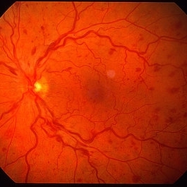
 Central Retinal Vein Occlusion: Case 1
Oct 12 2012 by Gregg T. Kokame, MD, MMM, FASRS
Color Fundus Photograph
Photographer: Jaclyn Pisano, Retina Consultants of Hawaii
Imaging device: Topcon 50IA / Eschalon
Condition/keywords: central retinal vein occlusion (CRVO)
-
 Non-Ischemic Central Retinal Vein Occlusion
Jun 28 2016 by Nichole Lewis
53-year-old female with non-ischemic central retinal vein occlusion left eye.
Photographer: Nichole Lewis
Condition/keywords: non-ischemic central retinal vein occlusion (CRVO)
 Loading…