Search results (104 results)
-
---thumb.JPG/image-square;max$300,300.ImageHandler) TID (Trans Illumination Defect)
TID (Trans Illumination Defect)
Jul 8 2013 by Jason S. Calhoun
74-year-old patient who VA 20/70 OD, 20/50 OS. Complaints of blurred vision. Iris atrophy, both eyes. ERM, right eye, Patient to have cataract surgery to improve distance vision.
Photographer: Jason S. Calhoun, Department of Ophthalmology, Mayo Clinic Jacksonville, Florida
Condition/keywords: translucency of iris
-
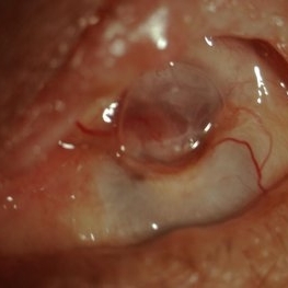
 Elschnig's Pearls
Sep 1 2015 by René Hernán Parada Vásquez
Fundus photograph of 58-year-old male with Elschnig's pearls, you can see the transparent clusters formed by proliferation of epithelial lens cells found in the remains of the capsule of the crystalline lens following cataract surgery.
Photographer: Parada René, ESO, Guatemala.
Condition/keywords: cataract surgery
-
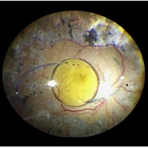
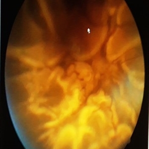 Closed Funnel Retinal Detachment
Apr 9 2017 by Aliya Sultana
Fundus phtograph of an 51-year-old man with closed funnel rhegmatogenous retinal detachment presented to our department 6 weeks after cataract surgery. Posterior capsule rent noticed with vitreous in anterior chamber, condensed vitreous tag is incarcerated in side port wound.
Photographer: Dr Aliya Sultana , Assistant Professor,Sarojini Devi Eye Hospital, Hyderabad, Telangana. India.
-
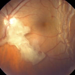
 Superior Peripapillary Hemorrhage
Jul 13 2013 by Jason S. Calhoun
Patient was seen for acute vision loss in the right eye. Patient has glaucoma. VA was 20/70 in the right eye. Had vitrectomy back in May 2012 for ERM stripping. Also had trabectome with cataract surgery in December of 2012. Fundus photos presents a superior peripapillary Hemorrhage of the optic nerve. Patient will be followed up in one month.
Photographer: Jason S. Calhoun, Department of Ophthalmology, Mayo Clinic Jacksonville, Florida
Imaging device: TOPCON TRC 50-EX
Condition/keywords: peripapillary hemorrhage
-
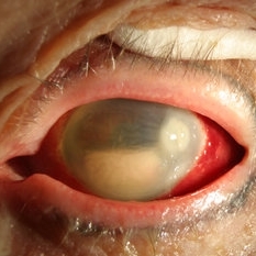
 Panophthalmitis
Jul 12 2014 by Philip J. Polkinghorne, MD
A 85-year-old lady who presented with an eroding intraocular lens. She had been initially treated with herbal medicines which failed to control the infection.
Photographer: Philip Polkinghorne
Condition/keywords: cataract surgery, endophthalmitis, panophthalmitis
-
 Superior Peripapillary Hemorrhage
Jun 27 2013 by Jason S. Calhoun
Patient was seen for acute vision loss in the right eye. Patient has glaucoma. VA was 20/70 in the right eye. Had vitrectomy back in May 2012 for ERM stripping. Also had trabectome with cataract surgery in December of 2012. Fundus photos presents a superior peripapillary Hemorrhage of the optic nerve. Patient will be followed up in one month.
Photographer: Jason S. Calhoun, Mayo Clinic Jacksonville, Florida
Imaging device: TOPCON TRC 50-EX
Condition/keywords: peripapillary hemorrhage
-
 Anterior Capsular Opacity
Feb 8 2018 by Claire Kiernan, MD
Slit lamp photograph of a 39-year-old female following uncomplicated cataract surgery shown here with dense fibrinous changes of the anterior capsule. This patient underwent Nd:YAG laser anterior capsulotomy with clearing of her visual axis.
Photographer: Steve Crow, University of Tennessee Hamilton Eye Institute, Memphis, TN
Condition/keywords: anterior capsule opacification, cataract extraction, cataract surgery
-
 Endophthalmitis
Apr 9 2014 by Aleksandra V. Rachitskaya, MD, FASRS
Slit lamp photo of a patient with endophthalmitis after cataract surgery. An infectious infiltrate is noted next to the clear corneal incision.
Photographer: Bascom Palmer Eye Institute
Condition/keywords: cataract surgery, endophthalmitis
-
---thumb.jpg/image-square;max$300,300.ImageHandler) Fundus Panorama: Post Operative Findings of Tractional Retinal Detachment
Fundus Panorama: Post Operative Findings of Tractional Retinal Detachment
Dec 25 2013 by Dong Yoon Kim, MD
47-year-old woman underwent vitrectomy and silicone oil tampoande for tractional retinal detachment due to proliferative diabetic retinopathy. 3 years after silicone oil removal and cataract surgery, her visual acuity was improved from hand motion to 20/40.
Condition/keywords: silicone oil, tractional retinal detachment
-
 Retained Lens Fragment
Mar 2 2014 by Homayoun Tabandeh, MD, FASRS
Retained lens fragment, choroidal detachment, and serous retinal detachment post cataract surgery
Condition/keywords: retained lens fragments
-
 OCT CME
Jul 9 2014 by Susanna S. Park, MD, PhD
OCT of a 72-year-old man with vision loss 6 months after cataract surgery showing CME.
Photographer: Ellen Redenbo
Condition/keywords: cystoid macular edema (CME)
-
 Pseudoexfoliation With Partial Subluxation
Jul 12 2013 by Jason S. Calhoun
81-year-old female woke up with loss of vision in the left eye. Patient was hand motion in the left eye. Slit lamp examination shows dislocated lens with pseudoexfoliation material around the iris rim. Proceeded with cataract surgery.
Photographer: Jason S. Calhoun, Department of Ophthalmology, Mayo Clinic Jacksonville, Florida
Condition/keywords: pseudoexfoliation of lens capsule, subluxation of lens
-
 Luxated IOL
Dec 16 2016 by Andrea Arriola-Lopez, MD MSc
56-year-old woman, PO posterior capsular rupture and sulcus IOL cataract surgery. Blurred vision, no pain. Found out a dropped IOL on a vitrectomized eye.
Photographer: Andrea Elizabeth Arriola-Lopez MD MSc
Imaging device: OR photo
Condition/keywords: dislocated intraocular lens (IOL), dislocated posterior chamber intraocular lens (PCIOL)
-
 Management of Ruptured Capsule During Cataract Surgery
Dec 10 2012 by Yale L. Fisher, MD
Dr. Steve Charles discussed his well researched approach to managing ruptured capsules experienced during cataract surgery. NOTE: A narration by Dr. Steve Charles will soon be available for this movie- please check back periodically.
Condition/keywords: cataract, ruptured capsules, video
-
 Retained Lens Fragments
Jul 13 2013 by Jason S. Calhoun
Post-op cataract surgery, slit lamp shows retained lens fragments in the lower portion on the anterior chamber.
Photographer: Jason S. Calhoun, Department of Ophthalmology, Mayo Clinic Jacksonville, Florida
Imaging device: TOPCON D-90 SL NIKON CAMERA
Condition/keywords: retained lens fragments
-
 Anterior Capsular Contraction Syndrome
Feb 8 2018 by Claire Kiernan, MD
Slit lamp photo of a female with underlying Retinitis Pigmentosa Zonulopathy and new anterior capsular contraction syndrome following cataract surgery.
Photographer: Steve Crow, University of Tennessee Hamilton Eye Institute, Memphis, TN
Condition/keywords: anterior capsule opacification, cataract surgery, zonules
-
 Subconjuntival IOL After Blunt Trauma
Jun 27 2018 by Gabriel Costa Andrade, PhD
A 73-year-old male patient was referred to our ophthalmic emergency department with complaints of redness, pain, and diminution of vision in his left eye, after fall from height. The patient underwent small incision cataract surgery with polymethylmethacrylate (PMMA) IOL implantation in both the eyes 15 years back through superior sclerocorneal incision under local anesthesia. His best-corrected visual acuity was perception of light in the left eye. Ophthalmic examination using slit lamp biomicroscopy of the left eye revealed diffuse subconjunctival hemorrhage with no conjunctival laceration and inferior bulbar conjunctiva showed traumatic pseudophacocele with a sign “golden half ring,” suggesting the presence of PCIOL in subconjunctival space.There was total hyphema obscuring the view of rest of the ocular structures in his left eye.
Photographer: Gabriel Andrade, RETINA CLINIC, São Paulo, BRAZIL
Condition/keywords: dislocated intraocular lens (IOL), trauma
-
 Dislocated IOL
May 15 2018 by Morgan Benton
Ultra-wide field pseudocolor image of a 68-year-old male with a dislocated IOL after cataract surgery in the left eye. Patient was only able to count fingers at one foot and could pinhole to 20/60.
Photographer: Morgan Benton
Imaging device: Optos
Condition/keywords: color fundus photograph, dislocated intraocular lens (IOL), left eye, Optos, ultra-wide field imaging
-
 SD-OCT of Ocular Hypotony
May 29 2013 by Zofia Anna Nawrocka (vel Michalewska), MD, PhD
SD-OCT of a 75-year-old patient with hypotony, 2 weeks after trauma, 2 years after extracapsular cataract surgery.
Photographer: Zofia Michalewska, Ophthalmic Clinic "Jasne Blonia
Imaging device: Spectralis
Condition/keywords: hypotony, optical coherence tomography (OCT)
-
 Fluorescein Angiography of Ocular Hypotony
May 29 2013 by Zofia Anna Nawrocka (vel Michalewska), MD, PhD
Fluorescein angiography of a 7- year-old patient with hypotony, 2 weeks after trauma, 2 years after extracapsular cataract surgery.
Photographer: Zofia Michalewska, Ophthalmic Clinic "Jasne Blonia
Imaging device: Spectralis
Condition/keywords: hypotony
-
 Spontaneous Dislocation of Capsular Bag
Jul 26 2015 by Mehul A Shah
A 57-year-old male presented with complaint of loss of vision after 3 years of cataract surgery, on examination his lens bag found dislocated in anterior chamber.
Photographer: Mehul Shah, Drashti Netralaya
Condition/keywords: intraocular lens dislocation
-
 AV Anastomosis
Sep 19 2017 by Purva Patwari
54-year-old female post cataract surgery.
Photographer: Dr Purva Patwari, Patwari Retina Center,Ahmedabad
Imaging device: Zeiss visu 500
Condition/keywords: arteriovenous anastomosis
-
 Scleritis
Jul 16 2014 by John S. King, MD
Keratoscleritis with suprachoroidal hemorrhage in an elderly male with history of cataract surgery.
Photographer: URMC
Condition/keywords: keratitis, scleritis, suprachoroidal hemorrhage
-
 Pseudoexfoliation With Partial Subluxation
Jul 12 2013 by Jason S. Calhoun
81-year-old female who woke up with loss of vision in the left eye. Patient was hand motion in the left eye. Slit lamp examination shows dislocated lens with pseudoexfoliation material around the iris rim. Proceeded with cataract surgery.
Photographer: Jason S. Calhoun, Department of Ophthalmology, Mayo Clinic Jacksonville, Florida
Condition/keywords: pseudoexfoliation of lens capsule, subluxation of lens
-
 Autofluorescence of Ocular Hypotony
May 29 2013 by Zofia Anna Nawrocka (vel Michalewska), MD, PhD
Autofluorescence image of a 75-year-old patient with hypotony, 2 weeks after trauma, 2 years after extracapsular cataract surgery.
Photographer: Zofia Michalewska, Ophthalmic Clinic "Jasne Blonia
Imaging device: Spectralis
Condition/keywords: hypotony
 Loading…