Search results (222 results)
-
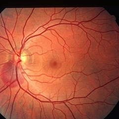
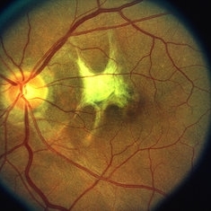
 Commotio Retinae
Mar 2 2014 by Homayoun Tabandeh, MD, FASRS
Commotio retinae following blunt trauma.
Condition/keywords: commotio retinae
-
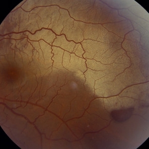
 Traumatic Retinal Dialysis-RD
Jan 1 2013 by John T. Thompson, MD
Traumatic retinal dialysis with localized retinal detachment after blunt trauma.
Condition/keywords: acute retinal detachment, retinal dialysis, retinal tear
-
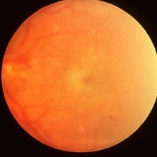
 Commotio retinae
Jan 11 2013 by Alex P. Hunyor, MD
Commotio retinae in the temporal mid periphery in an eye which sustained blunt trauma.
Condition/keywords: Berlin's edema, commotio retinae
-
 Traumatic Macular Hole
Aug 23 2012 by Gabriela Lopezcarasa Hernandez, MD
12-year-old boy with blunt trauma in right eye and central scotoma.
Photographer: Gabriela Lopezcarasa Hernandez, Hospital Angeles Lomas
Imaging device: ZEISS F4
Condition/keywords: blunt trauma, central scotoma, macular hole
-
 Traumatic Macular Hole
Aug 23 2012 by Gerardo Garcia-Aguirre, MD
Fundus photograph of a large macular hole with an area of pigment migration secondary to blunt trauma.
Photographer: Noemí Hernández, Asociación para Evitar la Ceguera en México
Imaging device: Zeiss FF4
Condition/keywords: deformity, macular hole
-
 Commotio retinae
Apr 29 2022 by Otakar Dušek, M.D. Ph.D.
Color fundus photograph of a 24-year-old woman who was hit by a volleyball in her right eye. This caused whitening of the lower peripheral retina (Berlin's edema) i.e. commotio retinae.
Photographer: Otakar Dušek, Charles University, Prague
Imaging device: Zeiss Clarus
Condition/keywords: Berlin's edema, blunt trauma, commotio retinae
-
 Juxtafoveal Choroidal Neovascularization Secondary to Choroidal Rupture
Aug 30 2012 by Young Hee Yoon, MD, PhD
SD-OCT image of a 14-year-old boy with a history of blunt trauma to his left eye 9 months ago. Best-corrected visual acuity remained at 20/30.
Photographer: Soo Hyun Cho, Asan Medical Center
Imaging device: HHeidelberg Spectralis OCTI/ version 1.7.0.0
Condition/keywords: choroidal rupture, juxtafoveal choroidal neovascularization (CNV)
-
 Juxtafoveal Choroidal Neovascularization Secondary to Choroidal Rupture
Aug 30 2012 by Young Hee Yoon, MD, PhD
Fundus photograph of a 14-year-old boy with a history of blunt trauma to his left eye 9 months ago. Best-corrected visual acuity remained at 20/30.
Photographer: Heon Eui Hong, Asan Medical Center
Imaging device: Canon CR-DGI / Version 5.1.2
Condition/keywords: choroidal rupture, juxtafoveal choroidal neovascularization (CNV)
-
 Berlin's Edema With Hemmorrhagic PED
Dec 12 2018 by Surendra Prakash, MBBS, MS, FELLOWSHIP IN VITREO RETINA
Fundus photograph of 24-year-old male having Berlin's edema with multiple sub RPE hemorrhage due to blunt trauma by football.
Photographer: DR SURENDRA PRAKASH
Condition/keywords: Berlin's edema, hemorrhagic detachment of retinal pigment epithelium
-
 Traumatic macular hole
Dec 19 2012 by Eric A. Postel, MD
Color fundus photograph of a young male with a traumatic macular hole
Condition/keywords: blunt trauma, macular hole
-
---thumb.jpg/image-square;max$300,300.ImageHandler) Commotio Retinae
Commotio Retinae
Jan 1 2013 by John T. Thompson, MD
commotio retinae after bullet blast near eye (non-perforating).
Condition/keywords: Berlin's edema, blunt trauma
-
 Retinitis Sclopetaria, 6 months later
Jun 29 2018 by Gareth Lema, MD, PhD
Retinitis sclopetaria has resolved. There are multiple, large choroidal ruptures and subretinal scarring.
Photographer: Flaum Eye Institute, University of Rochester, Rochester, NY
Condition/keywords: blunt trauma, chorioretinitis sclopetaria
-
 Intraocular Foreign Body, Human Eye Lash, Post Trauma - Right Eye
Oct 3 2012 by James B. Soque, CRA, OCT-C, COA, FOPS
30-year-old white male, with nail gun misfire and blunt force trauma to right eye. Nail penerated sup-nas globe OD, at the equator. Force of nail penetration, 'brought in' an Eye Lash Folicle. During vitrectomy, sterile FB OD was noted. Photograph captured 1 week post op. Color Fundus Photo, 50 Deg, Max 1X
Photographer: James Soque, CRA, COA, Island Retina, Shirley, NY, USA
Imaging device: Topcon TRC 50 DX, OIS Imaging Software
Condition/keywords: blunt trauma, intraocular foreign body, penetration
-
 Berlin's Edema
May 2 2013 by Henry J. Kaplan, MD
Macular Berlin's edema following blunt trauma.
Condition/keywords: Berlin's edema
-
 Post Traumatic Optic Nerve Head Avulsion
Nov 18 2017 by Vishal Agrawal, MD, FRCS,FACS,FASRS
Right eye fundus picture of a 24-year-old male patient who suffered blunt trauma 7 days back with a wooden stick . He presented with NLP vision with a non reacting dilated pupil. Fundus montage picture shows ONH avulsion,CRAO,peripapillary resolving hemorrhages and cicatricial tissue at the edge.
Photographer: Vishal Agrawal, MD, SMS Medical College, Jaipur, India
Imaging device: Zeiss 524
Condition/keywords: avulsion, central retinal artery occlusion (CRAO)
-
 Traumatic Peripapillary Hemorrhage
May 2 2013 by Henry J. Kaplan, MD
Peripapillary subretinal hemorrhage in the left eye after blunt trauma.
Condition/keywords: blunt trauma, peripapillary hemorrhage, subretinal hemorrhage
-
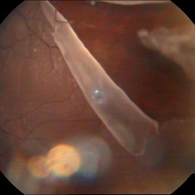
 Traumatic Lens Drop in Vitreous
Dec 15 2020 by Manish Nagpal, MD, FRCS (UK), FASRS
Patient had come to us status post blunt trauma with the lens dislocated in inferior vitreous.
Photographer: Gayathri Mohan, Retina Fellow, Retina Foundation, Ahmedabad, India
Imaging device: Mirante CSLO
Condition/keywords: dropped nucleus, lens dislocation, traumatic cataract
-
 Juxtafoveal Choroidal Neovascularization Secondary to Choroidal Rupture
Aug 30 2012 by Young Hee Yoon, MD, PhD
Fluorescence Angiography (FA) image of a 14-year-old boy with a history of blunt trauma to his left eye 9 months ago. Best-corrected visual acuity remained at 20/30.
Photographer: Heon Eui Hong, Asan Medical Center
Imaging device: HHeidelberg HRA II/ version 1.7.0.0
Condition/keywords: choroidal rupture, juxtafoveal choroidal neovascularization (CNV)
-
 Gonioscopy, Blood in the Anterior Chamber from Hyphema
Jul 8 2013 by Jason S. Calhoun
Patient with blunt trauma to the right eye due to a BB gun incident. Patient was present with a hyphema at 8-o'clock about 1mm thick. Gonioscopy photos were then taken to show blood from the hyphema entered into the anterior chamber. Patient had no angle recession in the right eye.
Photographer: Jason S. Calhoun, Department of Ophthalmology, Mayo Clinic Jacksonville, Florida
Condition/keywords: angle recession, gonioscopy
-
 Traumatic Retinal Tear
Sep 10 2014 by Mehul A Shah
A myopic male patient 30-years-old presented to outdoor and found to have retinal detachment with giant tear following blunt trauma
Photographer: Drashti Netralaya,Dahod
Imaging device: FF 450
Condition/keywords: giant retinal tear
-
 Traumatic Mac Scar
May 2 2013 by Henry J. Kaplan, MD
Traumatic stellate shaped subretinal macular scar.
Condition/keywords: blunt trauma, macular scar
-
 Ruptured Globe Trauma
Jul 11 2013 by Jason S. Calhoun
Young male who got hit with a baseball ended up with orbital floor fracture and ruptured globe.
Photographer: Jason S. Calhoun, Department of Ophthalmology, Mayo Clinic Jacksonville, Florida
Condition/keywords: blunt trauma
-
---thumb.jpg/image-square;max$300,300.ImageHandler) Traumatic Dislocation of Hypermature Cataract
Traumatic Dislocation of Hypermature Cataract
Jan 1 2013 by John T. Thompson, MD
Hypermature cataract with traumatic dislocation into anterior chamber.
Condition/keywords: anterior dislocation of lens, blunt trauma, hypermature cataract
-
 Berlin's Edema
Apr 8 2019 by Gary R. Cook, MD, FACS
39-year-old white female with geographic area of retinal whitening ( Berlin's edema) without hemorrhage in the midperiphery secondary to blunt trauma; V.A. = 20/25
Imaging device: Topcon VT-50
Condition/keywords: Berlin's edema, blunt trauma, retinal edema
-
 Juxtafoveal Choroidal Neovascularization Secondary to Choroidal Rupture
Aug 30 2012 by Young Hee Yoon, MD, PhD
Indocyanine Green Angiography (ICGA) image of a 14-year-old boy with a history of blunt trauma to his left eye 9 months ago. Best-corrected visual acuity remained at 20/30.
Photographer: Heon Eui Hong, Asan Medical Center
Imaging device: HHeidelberg HRA II/ version 1.7.0.0
Condition/keywords: choroidal rupture, juxtafoveal choroidal neovascularization (CNV)
 Loading…