Search results (266 results)
-
 Acute Posterior Multifocal Placoid Pigment Epitheliopathy
Sep 15 2012 by Roy D. Brod, MD
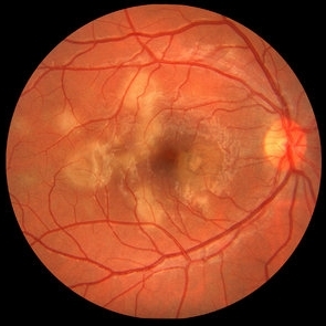
Fundus photograph right eye demonstrating cream colored placoid lesions in 28-year-old male patient with 4-day history of multiple scotomas OU.
Photographer: Julia Walker
Condition/keywords: acute posterior multifocal placoid pigment epitheliopathy (APMPPE), placoid retinal lesions, scotoma
-
 Acute Posterior Multifocal Placoid Pigment Epitheliopathy
Sep 15 2012 by Roy D. Brod, MD
Fundus photograph left eye demonstrating cream colored placoid lesions in 28-year-old male patient with 4-day history of multiple scotomas OU.
Photographer: Julia Walker
Condition/keywords: acute posterior multifocal placoid pigment epitheliopathy (APMPPE), placoid retinal lesions, scotoma
-
---thumb.jpg/image-square;max$300,300.ImageHandler) APMPPE Late Stage Scar Formation
APMPPE Late Stage Scar Formation
Feb 27 2013 by Henry J. Kaplan, MD
APMPPE late stage, multiple scar formation, left eye #2.
Condition/keywords: acute posterior multifocal placoid pigment epitheliopathy (APMPPE), late stage, white dot syndrome
-
 AMPPE
AMPPE
Oct 16 2012 by Ratimir Lazic, MD, PhD
Color fundus photography of a 58 -year-old male. Yellow- white placoid dots can be seen in macular region. BCVA of that eye is 0.9.
Photographer: Marko Lukic, MD
Imaging device: Zeis Visucam Lite 2
Condition/keywords: acute posterior multifocal placoid pigment epitheliopathy (APMPPE)
-
---thumb.jpg/image-square;max$300,300.ImageHandler) Acute Posterior Multifocal Placoid Pigment Epitheliopathy Late Stage Scar Formation
Acute Posterior Multifocal Placoid Pigment Epitheliopathy Late Stage Scar Formation
Feb 27 2013 by Henry J. Kaplan, MD
APMPPE late stage scar formation. Right Eye Multiple scar formations occurs in some of the patients #1
Condition/keywords: acute posterior multifocal placoid pigment epitheliopathy (APMPPE), late stage, white dot syndrome
-
 Acute Posterior Multifocal Placoid Pigment Epitheliopathy
Sep 15 2012 by Roy D. Brod, MD
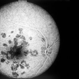
Late phase fluorescein angiogram demonstrating staining of placoid lesions in patient with APMPPE.
Photographer: Julia Walker
Condition/keywords: acute posterior multifocal placoid pigment epitheliopathy (APMPPE)
-
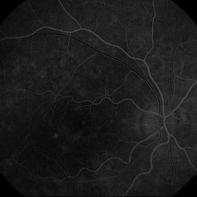
 AMPPE FA Early
Oct 9 2012 by Alan D. Letson, MD
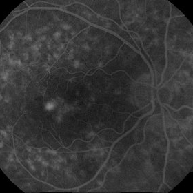
FA early
Photographer: Beverly Radcliffe
Condition/keywords: acute posterior multifocal placoid pigment epitheliopathy (APMPPE)
-
 Acute Multifocal Placoid Pigment Epitheliopathy Placoid Lesions
Oct 16 2012 by Jeffrey G. Gross, MD, FASRS
AMPPE placoid lesions, 20/15.
Condition/keywords: acute posterior multifocal placoid pigment epitheliopathy (APMPPE)
-
 APMPPE With Serous Macular Detachment
Jun 2 2014 by Rameez N Hussain, MD
Acute posterior multifocal placoid pigment epitheliopathy (APMPPE) with serous macular detachment.
Photographer: Rameez N Hussain MD, Vitreo Retinal Services, Giridhar Eye Institute, Cochin, India
Imaging device: Zeiss FF4
Condition/keywords: acute posterior multifocal placoid pigment epitheliopathy (APMPPE), serous retinal detachment
-
 CNV due to AMPPE
Oct 16 2012 by Ratimir Lazic, MD, PhD
Color fundus photography of a 58-year-old male. White dots with juxtafoveolar subretinal fluid can be seen. BCVA of that eye is 0.35.
Photographer: Marko Lukic, MD
Imaging device: Zeis Visucam Lite 2
Condition/keywords: acute posterior multifocal placoid pigment epitheliopathy (APMPPE), choroidal neovascularization (CNV)
-
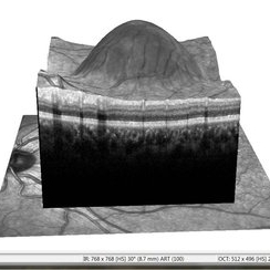
 APMPPE With Serous Macular Detachment SD-OCT
Jun 2 2014 by Rameez N Hussain, MD
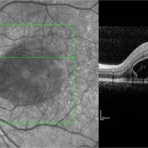
SD OCT image of acute posterior multifocal placoid pigment epitheliopathy (APMPPE) with serous macular detachment.
Photographer: Rameez N Hussain MD, Vitreo Retinal Services, Giridhar Eye Institute, Cochin, India
Imaging device: Heidelberg Spectralis
Condition/keywords: acute posterior multifocal placoid pigment epitheliopathy (APMPPE), serous retinal detachment
-
 CNV due to AMPPE
Oct 16 2012 by Ratimir Lazic, MD, PhD
FAG of 58-year-old male. In late venous phase hyperflorescence of white dots (caused by window defect) can be seen. Intensive leakage of dye in juxtafoveolar region.
Photographer: Marko Lukic, MD
Imaging device: Zeis Visucam Lite 2
Condition/keywords: acute posterior multifocal placoid pigment epitheliopathy (APMPPE), choroidal neovascularization (CNV)
-
---thumb.jpg/image-square;max$300,300.ImageHandler) Acute Posterior Multifocal Placoid Pigment Epitheliopathy
Acute Posterior Multifocal Placoid Pigment Epitheliopathy
Feb 27 2013 by Henry J. Kaplan, MD
APMPPE, fundus photographs. Left eye: Multiple placoid subretinal yellow - white lesions #2.
Condition/keywords: acute posterior multifocal placoid pigment epitheliopathy (APMPPE), white dot syndrome
-
 CNV due to AMPPE
Oct 16 2012 by Ratimir Lazic, MD, PhD
OCT image of 58-year- old male. Total resolution of fluid one and a half month after treatment can be seen. The patient was treated with intravitreal bevacizumab.
Photographer: Marko Lukic, MD
Imaging device: OCT Copernicus
Condition/keywords: acute posterior multifocal placoid pigment epitheliopathy (APMPPE), anti-VEGF, choroidal neovascularization (CNV)
-
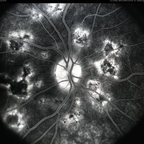
 AMPPE Fluorescein Angiogram late
Oct 9 2012 by Alan D. Letson, MD
FA, late in AMPPE patient
Photographer: Beverly Radcliffe
Condition/keywords: acute posterior multifocal placoid pigment epitheliopathy (APMPPE)
-
 APMPPE With Serous Macular Detachment 3D SD-OCT
Jun 2 2014 by Rameez N Hussain, MD
3D SD-OCT of acute posterior multifocal placoid pigment epitheliopathy (APMPPE) with serous macular detachment.
Photographer: Rameez N Hussain MD, Vitreo Retinal Services, Giridhar Eye Institute, Cochin, India
Imaging device: Heidelberg Spectralis
Condition/keywords: acute posterior multifocal placoid pigment epitheliopathy (APMPPE), serous retinal detachment
-
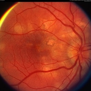
 Acute Multifocal Placoid Pigment Epitheliopathy
Oct 9 2012 by Alan D. Letson, MD
21-year-old femal college student presents with decreased vision OU after a viral prodrome.
Photographer: Beverly Radcliffe
Condition/keywords: acute posterior multifocal placoid pigment epitheliopathy (APMPPE)
-
---thumb.jpg/image-square;max$300,300.ImageHandler) APMPPE Late Stage Scar Formation
APMPPE Late Stage Scar Formation
Feb 27 2013 by Henry J. Kaplan, MD
APMPPE late stage scar formation. F/A hypofluorescence in the lesions area is due to masking effect of pigments . #1
Condition/keywords: acute posterior multifocal placoid pigment epitheliopathy (APMPPE), late stage, white dot syndrome
-
---thumb.jpg/image-square;max$300,300.ImageHandler) Acute Posterior Multifocal Placoid Pigment Epitheliopathy
Acute Posterior Multifocal Placoid Pigment Epitheliopathy
Feb 27 2013 by Henry J. Kaplan, MD
APMPPE. F/A .Late hyperfluorescence and staining of the lesions apparent #3.
Condition/keywords: acute posterior multifocal placoid pigment epitheliopathy (APMPPE), white dot syndrome
-
 CNV due to AMPPE
Oct 16 2012 by Ratimir Lazic, MD, PhD
OCT image of 58-year-old patient. Subfoveolar exudation and RPE defects can be seen. BCVA is 0.35.
Photographer: Marko Lukic, MD
Imaging device: SOCT Copernicus
Condition/keywords: acute posterior multifocal placoid pigment epitheliopathy (APMPPE), choroidal neovascularization (CNV)
-
---thumb.jpg/image-square;max$300,300.ImageHandler) AMPPE
AMPPE
Aug 12 2013 by From the Collections of Thomas M. Aaberg, MD and Thomas M. Aaberg Jr., MD
Chronic depigmentation.
Condition/keywords: acute posterior multifocal placoid pigment epitheliopathy (APMPPE), depigmentation
-
 CNV due to AMPPE
Oct 16 2012 by Ratimir Lazic, MD, PhD
FAG of 58-year-old male. In early venous phase hyperflorescence of white dots (caused by window defect) can be seen. Leakage of dye in juxtafoveolar region.
Photographer: Marko Lukic, MD
Imaging device: Zeis Visucam Lite 2
Condition/keywords: acute posterior multifocal placoid pigment epitheliopathy (APMPPE), choroidal neovascularization (CNV)
-
 APMPPE, Late Stage, Scar Formation
Mar 4 2013 by Henry J. Kaplan, MD
APMPPE, late stage, scar formation, F/A #2
Condition/keywords: acute posterior multifocal placoid pigment epitheliopathy (APMPPE), white dot syndrome
-
---thumb.jpg/image-square;max$300,300.ImageHandler) Acute Posterior Multifocal Placoid Pigment Epitheliopathy
Acute Posterior Multifocal Placoid Pigment Epitheliopathy
Feb 27 2013 by Henry J. Kaplan, MD
APMPPE fundus photographs. Right Eye multiple placoid yellowish subretinal lesions #1.
Condition/keywords: acute posterior multifocal placoid pigment epitheliopathy (APMPPE), white dot syndrome
-
 Acute Posterior Placoid Pigment Epitheliopathy
Mar 4 2013 by Henry J. Kaplan, MD
APMPPE; right eye; transition from acute stage to residual scar formation in some of the lesions. #1
Condition/keywords: acute posterior multifocal placoid pigment epitheliopathy (APMPPE), white dot syndrome
 Loading…