Search results (15 results)
-
 MNFL
MNFL
Sep 5 2015 by Ali Tavallali, MD, FASRS
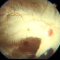
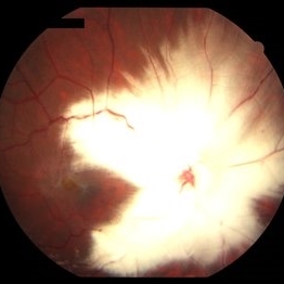
A 16-year-old malewith diffuse MNFL of OD.
Photographer: Neda Shaibani
Condition/keywords: myelinated nerve fiber layer
-
 MNFL
MNFL
Sep 5 2015 by Ali Tavallali, MD, FASRS
A 16-year-old male with diffuse MNFL of OD.
Photographer: Neda Shaibani
Condition/keywords: myelinated nerve fiber layer
-
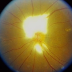
 Myelinated Nerve Fiber (mNFL)
Jun 21 2020 by Dhaivat Shah
Myelinated nerve fiber layer (mNFL) is a benign clinical entity that results from an embryologic developmental anomaly. Myelination along the visual pathway is noted around the eighth month of gestation, and typically reaches the posterior globe around the time of birth with virtually all fibers reaching complete myelination by age 7 months till the lamina cribrosa. Sometimes, due to altered neuro hormonal signals, this process of myelination extends past the lamina cribrosa and is visible on fundus examination as distinct white patches on the inner retinal surface. On infrared and red-free imaging, mNFL appears white, which is likely due to the high lipid content of myelin. Myelin blocks detection of underlying fluorescent material, thus appearing dark on fundus autofluorescence. On optical coherence tomography , it appears as a thickened and hyperreflective retinal nerve fiber layer. mNFL is typically benign but can be mistaken for other potentially serious conditions like neoplastic infiltration or infection. Hence, it is crucial to recognize the benign nature of mNFL to avoid superfluous medical testing.
Photographer: Ms Srishti Sharma
Imaging device: Choithram Netralaya
Condition/keywords: myelinated nerve fibers
-
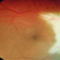
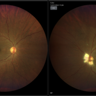
 Myelinated Nerve Fibre (MNF)
Jun 17 2023 by Harsh Vardhan Singh, MS
Fundus photograph of 32-year-old male having good best corrected visual acuity in both eyes with right eye having high myopia & MNF as incidental finding
Photographer: Dr Harsh Vardhan Singh, Assistant Professor, AIIMS, Guwahati
Condition/keywords: medullated nerve fibers, MNF, myelinated nerve fiber layer, myelinated nerve fibers, Nerve fiber layer arrangements, NFL
-
 Myelinated Nerve Fiber
May 5 2021 by Priya Rasipuram Chandrasekaran, MBBS, DO, DNB, FRCS
A 31-year-old male presented with a decreased vision of 20/125 N24 with -6.50 DS/-3.50 cyl 90 in the left eye. Fundus examination revealed peripapillary MNF progressing superiorly, obscuring disc and vessels and sparing the macula. OCT of ONH showed hyper reflective NFL and an abrupt ending of RPE and inner retinal layers (IRL) with underlying shadowing at the beginning of hyper reflectivity. The absence of photoreceptor integrity line (PIL) in the macula is believed to cause refractory amblyopia in such patients.
Condition/keywords: myelinated nerve fibers
-
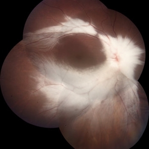
 Myelinated Nerve Fiber With Epiretinal Membrane With Lamellar Macular Hole
May 4 2020 by SWATI INDURKHYA
Heidelberg HRA + OCT Spectralis Multicolor (30 degree) retinal image of the left eye of a 28-year-old male showing myelinated nerve fibre (MNF) with epiretinal membrane (ERM) and lamellar macular hole (LMH).
Photographer: Rakesh PR, Giridhar Eye Institute, Kerala, India
Imaging device: Heidelberg Spectralis HRA + OCT
Condition/keywords: epiretinal membrane (ERM), myelinated nerve fibers
-
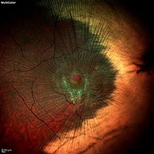
 MNF with ERM with LMH
Mar 24 2020 by Sugnesh Parmar
32-year-old male with unilateral high myopia with amblyopia on routine fundus examination was having large MNF with ERM with LMH
Photographer: Dr. Sugnesh Parmar, Radheshyam retina hospital, Bhavnagar, Gujarat, India
Condition/keywords: high myopia, lamellar macular hole
-
 Myelinated Nerve Fibres in left eye with old tributary vein occlusion in left eye
Jul 18 2023 by Harsh Vardhan Singh, MS
55 year female with left eye amblyopia & high myopia with MNF and Right eye showed signs of old macular branch retinal vein occlusion
Photographer: Harsh Vardhan Singh, AIIMS, Guwahati
Imaging device: Zeiss Clarus 700
Condition/keywords: BRVO, macular branch retinal vein occlusion (BRVO), Medullated Nerve fibres, MNF, Myelinated Nerve Fibres, TRVO
-
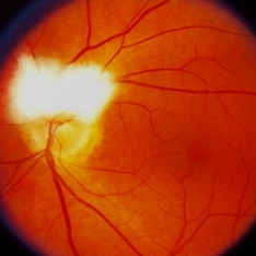
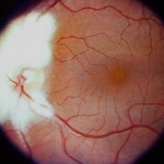 Myelinated Nerve Fibre (MNF)
Sep 26 2023 by Ben Serar
Fundus photograph of LE showing myelinated nerve fibres around the disc, sparing the nasal disc margin.
Condition/keywords: Myelinated Nerve Fibre (MNF)
-
 Myelinated Nerve Fibre (MNF)
Sep 12 2023 by Ben Serar
Fundus photograph of RE showing Myelinated Nerve Fibre along superior disc margin
Condition/keywords: MNF, myelinated nerve fiber
-
 Myelinated Nerve Fibres
Jan 30 2024 by Akansha Sharma
Color fundus photograph of a 15 year old male with myelinated nerve fibres all around the disc in the right eye.
Photographer: Dr. Akansha Sharma, Bharati Eye Hospital
Condition/keywords: MNF, myelinated nerve fibers
-
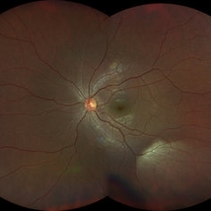 Myelinated Nerve Fibres
Jul 20 2023 by Harsh Vardhan Singh, MS
Fundus photograph of 32-year-old male with myelinated nerve fibre as incidental finding
Photographer: Dr Harsh Vardhan Singh, AIIMS, Guwahati
Imaging device: Zeiss Clarus 700
Condition/keywords: Medullated Nerve fibres, MNF, Myelinated Nerve Fibres
-
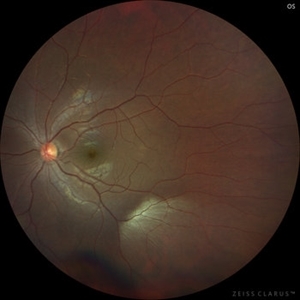 Myelinated Nerve Fibres
Jul 20 2023 by Harsh Vardhan Singh, MS
Fundus photograph of 32-year-old male with myelinated nerve fibre as incidental finding
Photographer: Dr Harsh Vardhan Singh, AIIMS, Guwahati
Imaging device: Zeiss Clarus 700
Condition/keywords: Medullated Nerve fibres, MNF, Myelinated Nerve Fibres
-
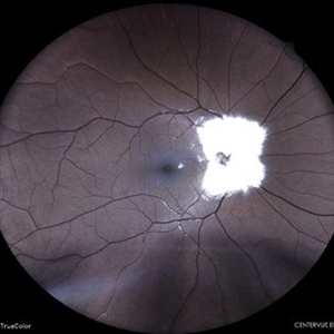
 Myelinated Nerve Fibre (MNF)
Sep 21 2023 by Ben Serar
Fundus photograph showing myelinated nerve fibre superior to the disc obscuring the superior disc margin, and also inferior to disc.
Condition/keywords: Myelinated Nerve Fibre (MNF)
-
 Myelinated Nerve Fibre (MNF)
Sep 12 2023 by Ben Serar
Fundus photograph of LE showing Myelinated Nerve Fibre along superior disc margin.
Condition/keywords: Myelinated Nerve Fibre (MNF)
 Loading…