Search results (309 results)
-
 Acute Posterior Multifocal Placoid Pigment Epitheliopathy
Feb 20 2024 by Soobien Lee
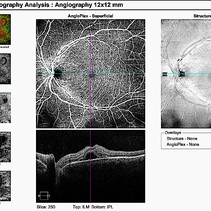
12x12mm OCT Angiography of a 20-year-old caucasian female with viral prodrome and vision loss OS>OD secondary to Acute Posterior Multifocal Placoid Pigment Epitheliopathy (APPME). Imaging shows multifocal flow voids.
Photographer: Kim Seay, Elman Retina Group
Imaging device: 12x12mm OCT-Angiography
Condition/keywords: acute posterior multifocal placoid pigment epitheliopathy (APMPPE), bacillary layer detachment, OCT, OCT Angiography, Uveitis, white dot syndrome
-
 Acute Posterior Multifocal Placoid Pigment Epitheliopathy
Feb 20 2024 by Soobien Lee
Optos color fundus photograph of a 20-year-old caucasian female with viral prodrome and vision loss OS>OD secondary to Acute Posterior Multifocal Placoid Pigment Epitheliopathy (APPME). Imaging of her left eye shows multiple bilateral creamy yellow-white placoid lesions at the level of RPE and choroid throughout the posterior pole.
Photographer: Ashley Metzger, Elman Retina Group
Imaging device: Optos Ultra-Widefield Imaging
Condition/keywords: acute posterior multifocal placoid pigment epitheliopathy (APMPPE), bacilliary layer detachment, Optos, uveitis, white dot syndrome
-
 Acute Posterior Multifocal Placoid Pigment Epitheliopathy
Feb 20 2024 by Soobien Lee
Optos fundus autofluorescence photograph of a 20-year-old caucasian female with viral prodrome and vision loss OS>OD secondary to Acute Posterior Multifocal Placoid Pigment Epitheliopathy (APPME). Imaging of her left eye shows hypoautofluorescent areas corresponding to multiple bilateral placoid lesions at the level of RPE and choroid throughout the posterior pole.
Photographer: Ashley Metzger, Elman Retina Group
Imaging device: Optos Ultra-Widefield Autoflurescence Imaging
Condition/keywords: acute posterior multifocal placoid pigment epitheliopathy (APMPPE), autofluorescence imaging, bacilliary layer detachment, Optos, OPTOS CALIFORNIA, uveitis, white dot syndrome
-
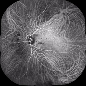
 Acute Posterior Multifocal Placoid Pigment Epitheliopathy
Feb 20 2024 by Soobien Lee
Fluorescein angiogram of a 20-year-old caucasian female with viral prodrome and vision loss OS>OD secondary to Acute Posterior Multifocal Placoid Pigment Epitheliopathy (APPME). Early blockage with late hyperfluorescent leakage can be seen on fluorescein angiography of the left eye.
Photographer: Ashley Metzger, Elman Retina Group
Imaging device: Optos Ultra-Widefield Fluorescein Angiography
Condition/keywords: acute posterior multifocal placoid pigment epitheliopathy (APMPPE), bacilliary layer detachment, FA, FA early phase, fluorescein angiogram (FA), Optos, uveitis, white dot syndrome
-
 Acute Posterior Multifocal Placoid Pigment Epitheliopathy
Feb 20 2024 by Soobien Lee
Fluorescein angiogram of a 20-year-old caucasian female with viral prodrome and vision loss OS>OD secondary to Acute Posterior Multifocal Placoid Pigment Epitheliopathy (APPME). Early blockage with late hyperfluorescent leakage can be seen on fluorescein angiography of the left eye.
Photographer: Ashley Metzger, Elman Retina Group
Imaging device: Optos Ultra-Widefield Fluorescein Angiography
Condition/keywords: acute posterior multifocal placoid pigment epitheliopathy (APMPPE), bacilliary layer detachment, FA, FA late phase, FA late phase leakage, fluorescein angiogram (FA), Optos, uveitis, white dot syndrome
-
 Acute Posterior Multifocal Placoid Pigment Epitheliopathy
Feb 20 2024 by Soobien Lee
A 20-year-old caucasian female with viral prodrome and vision loss OS>OD secondary to Acute Posterior Multifocal Placoid Pigment Epitheliopathy (APPME). OCT of the left macula shows bacillary layer detachment.
Photographer: Kim Seay, Elman Retina Group
Condition/keywords: acute posterior multifocal placoid pigment epitheliopathy (APMPPE), bacilliary layer detachment, OCT, Uveitis, white dot syndrome
-
---thumb.jpg/image-square;max$300,300.ImageHandler) APMPPE Late Stage Scar Formation
APMPPE Late Stage Scar Formation
Feb 27 2013 by Henry J. Kaplan, MD
APMPPE late stage scar formation. F/A hypofluorescence in the lesions area is due to masking effect of pigments . #1
Condition/keywords: acute posterior multifocal placoid pigment epitheliopathy (APMPPE), late stage, white dot syndrome
-
 MEWDS
MEWDS
Oct 9 2020 by David L Kilpatrick, MD
26-year-old female presented with unilateral vision loss. She c/o flashes and a peripheral scotoma. Vision was 20/100. On exam, she showed foveal granularity, mild disc edema, and white dots as seen. Three weeks later, white dots had resolved and vision improved to 20/25.
Photographer: Mississippi Retina Associates
Imaging device: Optos
Condition/keywords: multiple evanescent white dot syndrome (MEWDS)
-
 Serpiginous Choroiditis
Sep 22 2019 by Haider Ali
35-year-old female presented with decrease in vision in her left eye for last 4 days and in right eye for last 8 days. Her right eye was previously involved in a similar episode about 5-6 months ago for which she was treated with oral steroids.
Photographer: Dr Haider Ali Chaudhry, Madinah Teaching Hospital, Faisalabad
Condition/keywords: acute posterior multifocal placoid pigment epitheliopathy (APMPPE), macula serpiginous choroidopathy, posterior uveitis, serpiginous choroiditis, uveitis, white dot lesions, white dot syndrome
-
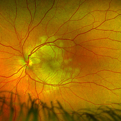
 Multifocal Choroiditis
Aug 16 2018 by FELIPE PEREIRA
Mid-phase indocyanine green angiography of a 25-year-old woman with sudden central vision loss and photopsias for 7 days. The hypofluorescent lesions in the macula and nasal to the disc correspond to the yellow-white deep lesions in the fundus examination. No leakage is observed at any stage of the exam
Photographer: Claudio Zett Lobos
Imaging device: HEIDELBERG SPECTRALIS HRA
Condition/keywords: indocyanine green (ICG) angiography, multifocal choroiditis, white dot syndrome
-
 Multiple evanescent White Dot Syndrome (MEWDS)
May 27 2025 by César Adrián Gómez Valdivia, MD
Fundus photograph of a 21 year-old female patient with suspected Multiple Evanescent White Dot Syndrome (MEWDS). The White Dot Syndromes produce yellow-white retinal lesions classically located at the retinal pigment epithelium or outer retina and are found primarily in young adults. Symptoms of MEWDS include unilateral blurred vision, visual field loss, photopsias, and floaters.
Photographer: @eyemissu2
Imaging device: TOPCON TRC-50DX
Condition/keywords: multiple evanescent white dot syndrome (MEWDS)
-
 Serpiginous Choroiditis
Sep 22 2019 by Haider Ali
35-year-old female presented with decrease in vision in her left eye for last 4 days and in right eye for last 8 days. Her right eye was previously involved in a similar episode about 5-6 months ago for which she was treated with oral steroids.
Photographer: Dr Haider Ali Chaudhry, Madinah Teaching Hospital, Faisalabad
Condition/keywords: acute posterior multifocal placoid pigment epitheliopathy (APMPPE), macula serpiginous choroidopathy, posterior uveitis, serpiginous choroiditis, uveitis, white dot lesions, white dot syndrome
-
 Serpiginous Choroiditis (Recurrent)
Sep 22 2019 by Haider Ali
35-year-old female presented with decrease in vision in her left eye for last 4 days and in right eye for last 8 days. Her right eye was previously involved in a similar episode about 5-6 months ago for which she was treated with oral steroids.
Photographer: Dr Haider Ali Chaudhry, Madinah Teaching Hospital, Faisalabad
Condition/keywords: acute posterior multifocal placoid pigment epitheliopathy (APMPPE), macula serpiginous choroidopathy, serpiginous choroiditis, white dot syndrome
-
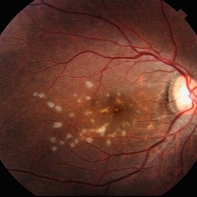
 White Dot Syndrome
Jan 11 2013 by Hyung-Woo Kwak, MD
The fundus showed multiple scattered white spots
Photographer: Misook Lee, Kyung Hee Univsersity Hospital, Seoul
Imaging device: Zeiss f 450 plus
Condition/keywords: white dot syndrome
-
---thumb.jpg/image-square;max$300,300.ImageHandler) Acute Posterior Multifocal Placoid Pigment Epitheliopathy
Acute Posterior Multifocal Placoid Pigment Epitheliopathy
Feb 27 2013 by Henry J. Kaplan, MD
APMPPE fundus photographs. Right Eye multiple placoid yellowish subretinal lesions #1.
Condition/keywords: acute posterior multifocal placoid pigment epitheliopathy (APMPPE), white dot syndrome
-
---thumb.jpg/image-square;max$300,300.ImageHandler) Acute Posterior Multifocal Placoid Pigment Epitheliopathy
Acute Posterior Multifocal Placoid Pigment Epitheliopathy
Feb 27 2013 by Henry J. Kaplan, MD
APMPPE, fundus photographs. Left eye: Multiple placoid subretinal yellow - white lesions #2.
Condition/keywords: acute posterior multifocal placoid pigment epitheliopathy (APMPPE), white dot syndrome
-
---thumb.jpg/image-square;max$300,300.ImageHandler) APMPPE Late Stage Scar Formation
APMPPE Late Stage Scar Formation
Feb 27 2013 by Henry J. Kaplan, MD
APMPPE late stage, multiple scar formation, left eye #2.
Condition/keywords: acute posterior multifocal placoid pigment epitheliopathy (APMPPE), late stage, white dot syndrome
-
---thumb.jpg/image-square;max$300,300.ImageHandler) Acute Posterior Multifocal Placoid Pigment Epitheliopathy Late Stage Scar Formation
Acute Posterior Multifocal Placoid Pigment Epitheliopathy Late Stage Scar Formation
Feb 27 2013 by Henry J. Kaplan, MD
APMPPE late stage scar formation. Right Eye Multiple scar formations occurs in some of the patients #1
Condition/keywords: acute posterior multifocal placoid pigment epitheliopathy (APMPPE), late stage, white dot syndrome
-
---thumb.jpg/image-square;max$300,300.ImageHandler) Acute Posterior Multifocal Placoid Pigment Epitheliopathy
Acute Posterior Multifocal Placoid Pigment Epitheliopathy
Feb 27 2013 by Henry J. Kaplan, MD
APMPPE, red free imaging: right eye #1.
Condition/keywords: acute posterior multifocal placoid pigment epitheliopathy (APMPPE), white dot syndrome
-
---thumb.jpg/image-square;max$300,300.ImageHandler) Acute Posterior Multifocal Placoid Pigment Epitheliopathy
Acute Posterior Multifocal Placoid Pigment Epitheliopathy
Feb 27 2013 by Henry J. Kaplan, MD
APMPPE. F/A .Late hyperfluorescence and staining of the lesions apparent #3.
Condition/keywords: acute posterior multifocal placoid pigment epitheliopathy (APMPPE), white dot syndrome
-
---thumb.jpg/image-square;max$300,300.ImageHandler) Birdshot Retinochoroidopathy
Birdshot Retinochoroidopathy
Feb 26 2013 by Henry J. Kaplan, MD
Birdshot retinochoroidopathy.
Condition/keywords: birdshot retinochoroidopathy, white dot syndrome
-
 Birdshot: a View From the Outside
Nov 3 2019 by Julia Farah, MD
61-year-old female presented with classic birdshot chorioretinopathy.
Photographer: Peter Guingab
Imaging device: Optos California
Condition/keywords: birdshot choroidopathy, uveitis, white dot syndrome
-
 MEWDS
MEWDS
Feb 14 2018 by Daniel K Bennett, MD
24-year-old white female with mild scotoma.
Imaging device: Zeiss Visucam
Condition/keywords: multiple evanescent white dot syndrome (MEWDS)
-
 Multiple Evanescent White Dot Syndrome (MEWDS)
Oct 20 2012 by Hyung-Woo Kwak, MD
Numerous small deep ill-defined, grey-white dot were seen at the posterior pole and mid-periphery. Some lesions showed mild hyperfluorescence in autofluorescence (AF) but were of limited diagnostic value. ICG showed more numerous hypofluorescent spots than are apparent clinically or on AF/FA
Condition/keywords: hypofluorescent spots, multiple evanescent white dot syndrome (MEWDS)
 Loading…