Search results (37 results)
-
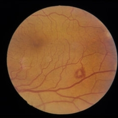
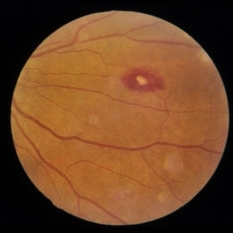
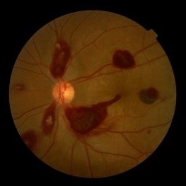 Ocular Manifestation of Acute Leukemia
Sep 8 2012 by Hamid Ahmadieh, MD
Color fundus photograph of a 26-year-old man with acute leukemia.
Photographer: Hamid Ahmadieh, MD, Ophthalmic Research Center, Labbafinejad Medical Center, Shahid Beheshti University of Medical Sciences , Tehran
Imaging device: Topcon Fundus Camera
Condition/keywords: acute leukemia, white centered retinal hemorrhage (Roth Spot)
-
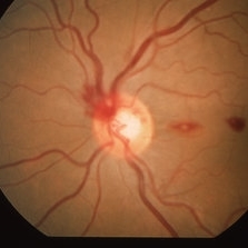
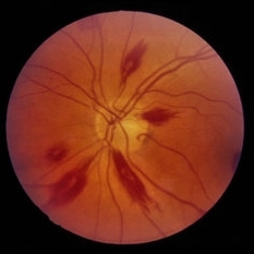
 Leukemic Retinopathy
Oct 9 2012 by Sharon Fekrat, MD FACS FASRS
22-year-old female with new diagnosis of acute myelogenous leukemia. White blood cell count was 35,000,000,000 cells/L. Note Roth Spots.
Photographer: Tiffanie Keaton, Duke Eye Imaging, Durham, NC
Condition/keywords: acute leukemia, white centered retinal hemorrhage (Roth Spot)
-
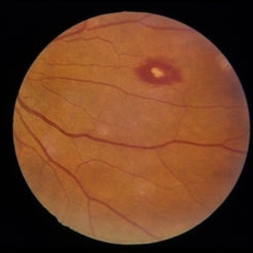
 Roth Spot
Dec 22 2014 by H. Michael Lambert, MD
White centered heme.
Condition/keywords: white centered retinal hemorrhage (Roth Spot)
-
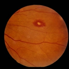
 Roth Spot
Oct 2 2013 by Jerald A. Bovino, MD
There is a white centered hemorrhage known as a Roth spot.
Condition/keywords: white centered retinal hemorrhage (Roth Spot)
-
 Thrombocytopenia
Sep 24 2024 by DR Rohit Gupta
Fundus photography of a 16 year-old girl suffering from severe thrombocytopenia, showing flame shaped hemorrhage.
Photographer: Dr Rohit gupta
Imaging device: Samsung S21
Condition/keywords: anaemic retinopathy, flame shaped retinal hemorrhage, Haemorrhage, Roth spots, white centered retinal hemorrhage (Roth Spot), white dot syndrome
-
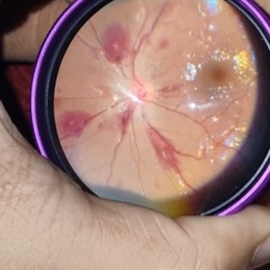
---thumb.jpg/image-square;max$300,300.ImageHandler) Roth Spot
Roth Spot
Feb 27 2013 by Henry J. Kaplan, MD
Roth spot due to subacute bacterial endocardiris in AIDS patient. Magnified view in the same patient.
Condition/keywords: AIDS, subacute bacterial endocardiris, white centered retinal hemorrhage (Roth Spot)
-
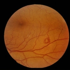
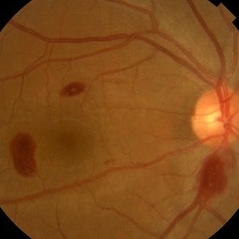
 Anemia
Mar 26 2019 by Gary R. Cook, MD, FACS
Retinal hemorrhages OS in a 45 year old female secondary to iron deficiency anemia; VA= 20/20.
Imaging device: Topcon VT-50
Condition/keywords: anemia, blot hemorrhages, hemorrhage, white centered retinal hemorrhage (Roth Spot)
-
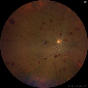
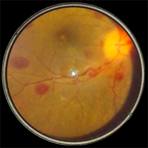
 Leukemic Retinopathy
Nov 27 2024 by Ramses Rosales-Diaz
Fundus photograph of a 48-year-old woman with venous dilatation and tortuosity, flame-shaped and intraretinal hemorrhages, Roth spots and sub-ILM hemorrhage. Her complete blood count reports 425,540 lymphocytes/microliter, and the blood smear reveals Gumprecht shadows and numerous lymphocytes with nuclei exhibiting hypercondensed chromatin. She is diagnosed with chronic lymphocytic leukemia and receives appropriate treatment from the hematology team
Photographer: Ramses Rosales-Diaz, Asociación Para Evitar la Ceguera en México
Imaging device: Clarus 700
Condition/keywords: leukemia, sub ILM hemorrhage, white centered retinal hemorrhage (Roth Spot)
-
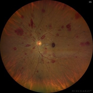
 Leukemic Retinopathy
Nov 27 2024 by Ramses Rosales-Diaz
Fundus photograph of a 48-year-old woman showing venous dilatation and tortuosity, flame-shaped hemorrhages, intraretinal hemorrhages, sub-ILM hemorrhages, and Roth spots. Her complete blood count shows 425,540 lymphocytes/microliter, and the blood smear reveals Gumprecht shadows along with numerous lymphocytes with hypercondensed chromatin in their nuclei. She is diagnosed with chronic lymphocytic leukemia and receives appropriate treatment from the hematology team.
Photographer: Ramses Rosales-Diaz, Asociación Para Evitar la Ceguera en México
Imaging device: Zeiss Clarus 700
Condition/keywords: leukemia, sub ILM hemorrhage, white centered retinal hemorrhage (Roth Spot)
-
 Leukemic Retinopathy
May 13 2014 by ayesha tasneem
Fundus photograph of a 25-year-old female patient with acute myeloblastic leukemia presenting with extensive white centered hemorrhages,and decreased vision.
Condition/keywords: white centered retinal hemorrhage (Roth Spot)
-
 Ocular Manifestation of Acute Leukemia
Sep 5 2012 by Hamid Ahmadieh, MD
Color fundus photograph of a 26-year-old man with acute leukemia.
Photographer: Hamid Ahmadieh, MD, Ophthalmic Research Center, Labbafinejad Medical Center, Shahid Beheshti University of Medical Sciences
Imaging device: Topcon Fundus Camera
Condition/keywords: acute leukemia, white centered retinal hemorrhage (Roth Spot)
-
---thumb.jpg/image-square;max$300,300.ImageHandler) Roth Spot
Roth Spot
Feb 27 2013 by Henry J. Kaplan, MD
Roth spots due to subacute bacterial endocardiris in a patient with the diagnosis of AIDS .
Condition/keywords: AIDS, subacute bacterial endocardiris, white centered retinal hemorrhage (Roth Spot)
-
 Roth Spot
Dec 22 2014 by H. Michael Lambert, MD
Looks more like a ring of small retinal hemorrhages.
Condition/keywords: white centered retinal hemorrhage (Roth Spot)
-
 Roth Spot
Dec 22 2014 by H. Michael Lambert, MD
Looks more like a ring of small retinal hemorrhages.
Condition/keywords: white centered retinal hemorrhage (Roth Spot)
-
 Roth Spot
Dec 22 2014 by H. Michael Lambert, MD
Numerous flame hemorrhages, several with white centers.
Condition/keywords: white centered retinal hemorrhage (Roth Spot)
-
 Roth Spot
Dec 22 2014 by H. Michael Lambert, MD
White centered heme.
Condition/keywords: white centered retinal hemorrhage (Roth Spot)
-
 Roth Spot
Dec 22 2014 by H. Michael Lambert, MD
White centered heme.
Condition/keywords: white centered retinal hemorrhage (Roth Spot)
-
 Roth Spots
Jul 11 2013 by Jerald A. Bovino, MD
No history.
Condition/keywords: white centered retinal hemorrhage (Roth Spot)
-
 Roth Spots
Jul 11 2013 by Jerald A. Bovino, MD
No history, part of stereo pair.
Condition/keywords: stereo pair, white centered retinal hemorrhage (Roth Spot)
-
 Roth Spots - Bacterial Endocarditis
Dec 3 2017 by John S. King, MD
Initial presentation; 29-year-old white female denied ivdu p/c with acute scotoma due to the sub-ILM foveal heme. She did have some roth spots in both eyes. There was a focal area of periphlebitis just superior to the fovea OD. Work up for roth spots and retinal vasculitis initiated. She did have a low grade fever that she attributed to a urinary tract infection being treated by her PCP.
Imaging device: Optos
Condition/keywords: sub-inner limiting membrane hemorrhage, white centered retinal hemorrhage (Roth Spot)
-
 Roth Spots - Bacterial Endocarditis
Dec 3 2017 by John S. King, MD
Few weeks later; some areas resolved, and new roth spots; sub-ILM foveal heme in right eye resolved and vision back to baseline; full work-up pending.
Imaging device: Optos
Condition/keywords: bacterial endocarditis, white centered retinal hemorrhage (Roth Spot)
-
 Roth Spots - Bacterial Endocarditis
Dec 3 2017 by John S. King, MD
Two months later; interim was hospitalized for bacterial infection of mitral valve; roth spots resolved.
Imaging device: Optos
Condition/keywords: bacterial endocarditis, white centered retinal hemorrhage (Roth Spot)
-
 Roth Spots : Smartphone Fundus Image
Dec 14 2018 by Prithvi Chandrakanth
A 13-year-old female presented with multiple white centered retinal hemorrhage in both the eyes.
Photographer: Dr.Prithvi Chandrakanth, Dr.Chandrakanth Malabar Nethralaya, Kozhikode.
Imaging device: Trash To Treasure Retcam : Smartphone Fundus Camera
Condition/keywords: Roth spots, smartphone fundus photography, white centered retinal hemorrhage (Roth Spot)
-
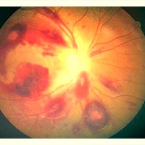
---thumb.jpg/image-square;max$300,300.ImageHandler) Roth Spots and Retinal Hemorrhage
Roth Spots and Retinal Hemorrhage
Dec 27 2013 by David Callanan, MD
24-year-old patient, AML/ post-chemo thrombocytopenia with pre, intra, & sub-retinal.
Condition/keywords: white centered retinal hemorrhage (Roth Spot)
 Loading…